미국/글로벌: +1-949-461-9292
EUROPE: +39-011-3052-794
문의하기

카테고리: 프로파일 측정 테스트


stent coating adhesion testing failure analysis drug eluting stent coating

Stent Coating Adhesion and Delamination Analysis Using Nano Scratch Testing

Application Note | Stent Coating Adhesion Testing

Stent Coating Adhesion and Delamination Analysis Using Nano Scratch Testing

Quantifying Coating Failure and Adhesion Performance on Drug-Eluting Stents

stent coating adhesion testing nano scratch delamination critical load

Research & Experimental Testing

Duanjie Li, PhD

Visual Design & Editorial

앤드류 쇼어

소개

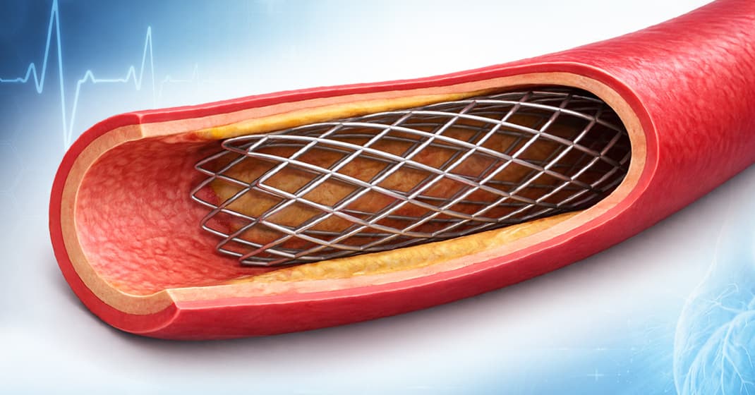
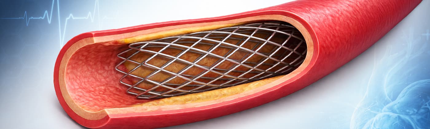
Blood is carried through arteries from the heart to the rest of the body. Any weakening or blockage of these vessels can pose significant health risks and may become life-threatening. A stent is a small mesh tube inserted into the lumen of a blood vessel to treat narrowed or weakened arteries. Stent implantation is now a widely used procedure to support the arterial wall and restore blood flowᶦ.

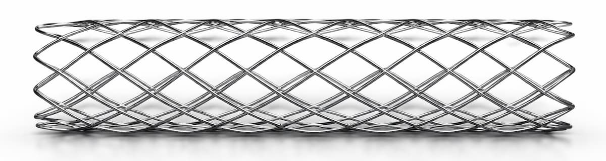
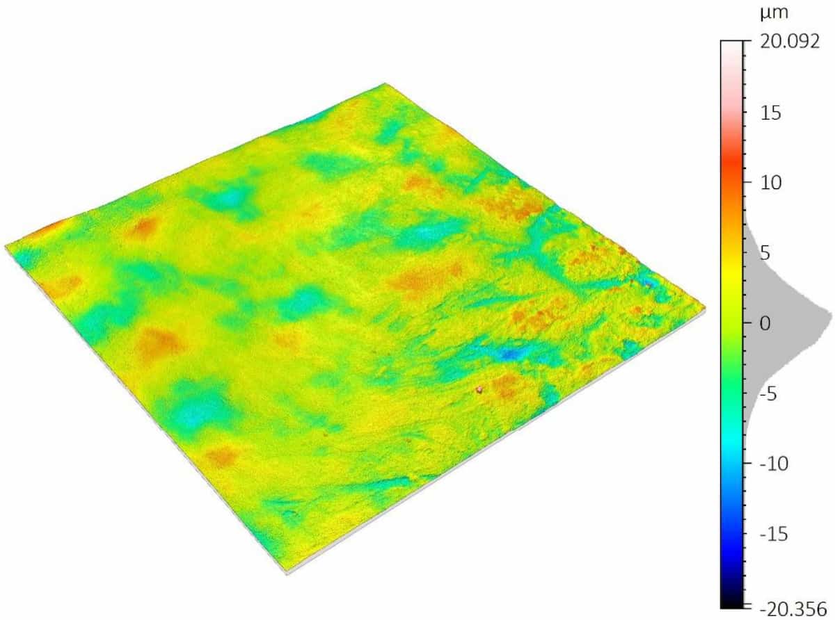
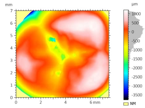
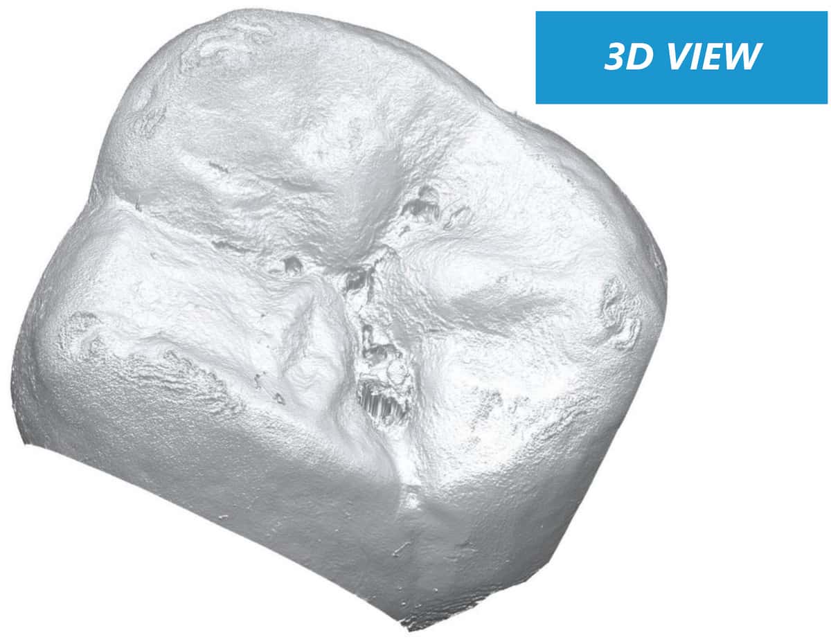
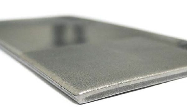
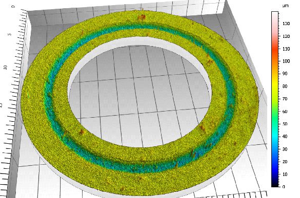
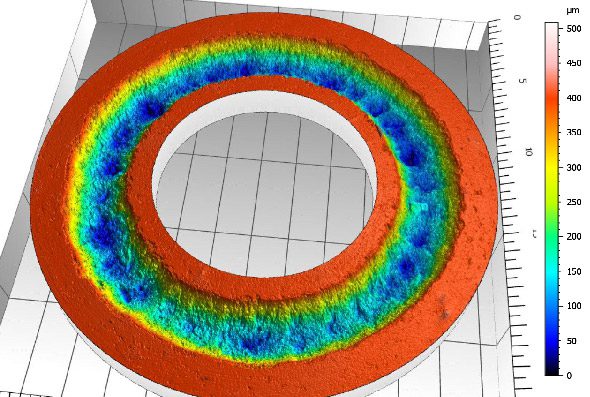
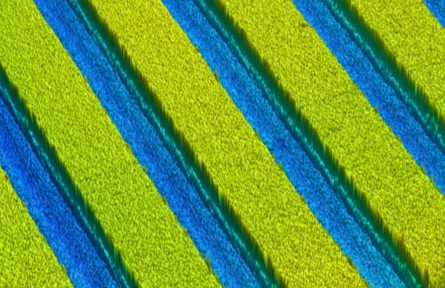
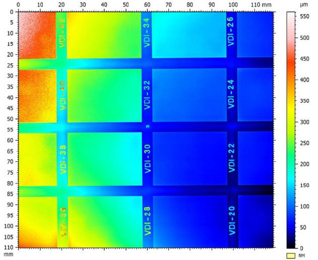
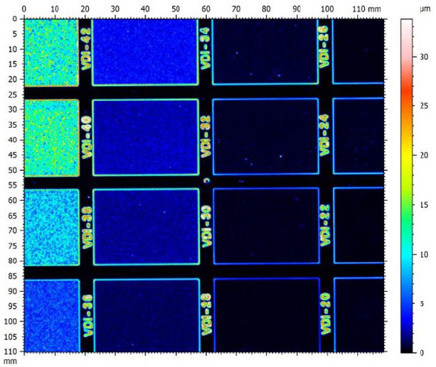
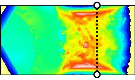
Metal stent mesh geometry illustrating the structural complexity of vascular implant design.

Why coating adhesion matters in drug-eluting stents

Drug-eluting stents represent a major advancement in stent technology. They incorporate a biodegradable, biocompatible polymer coating that enables controlled drug release at the arterial site, helping to inhibit intimal thickening and reduce the risk of restenosisᶦᶦ.

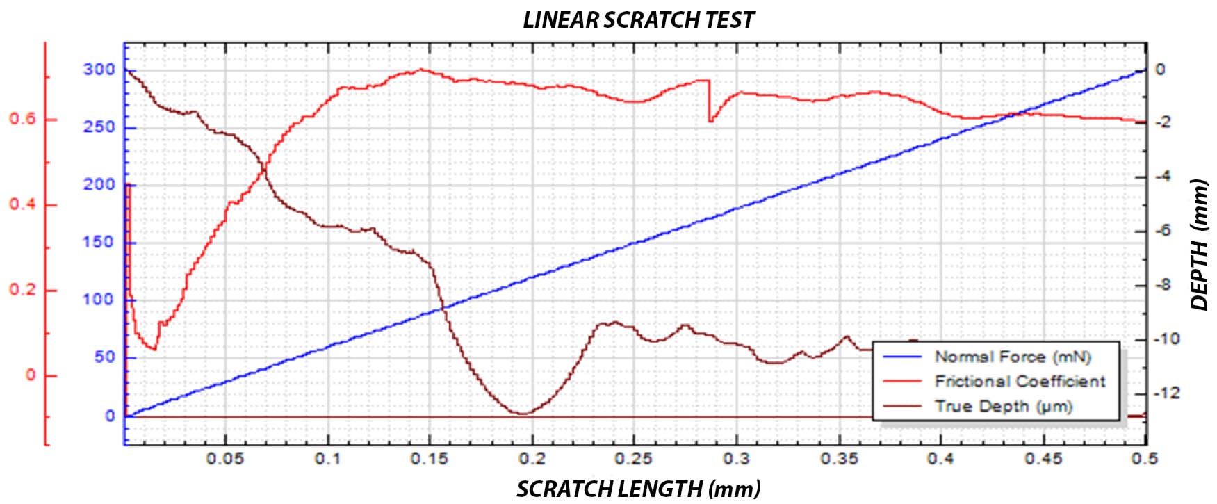
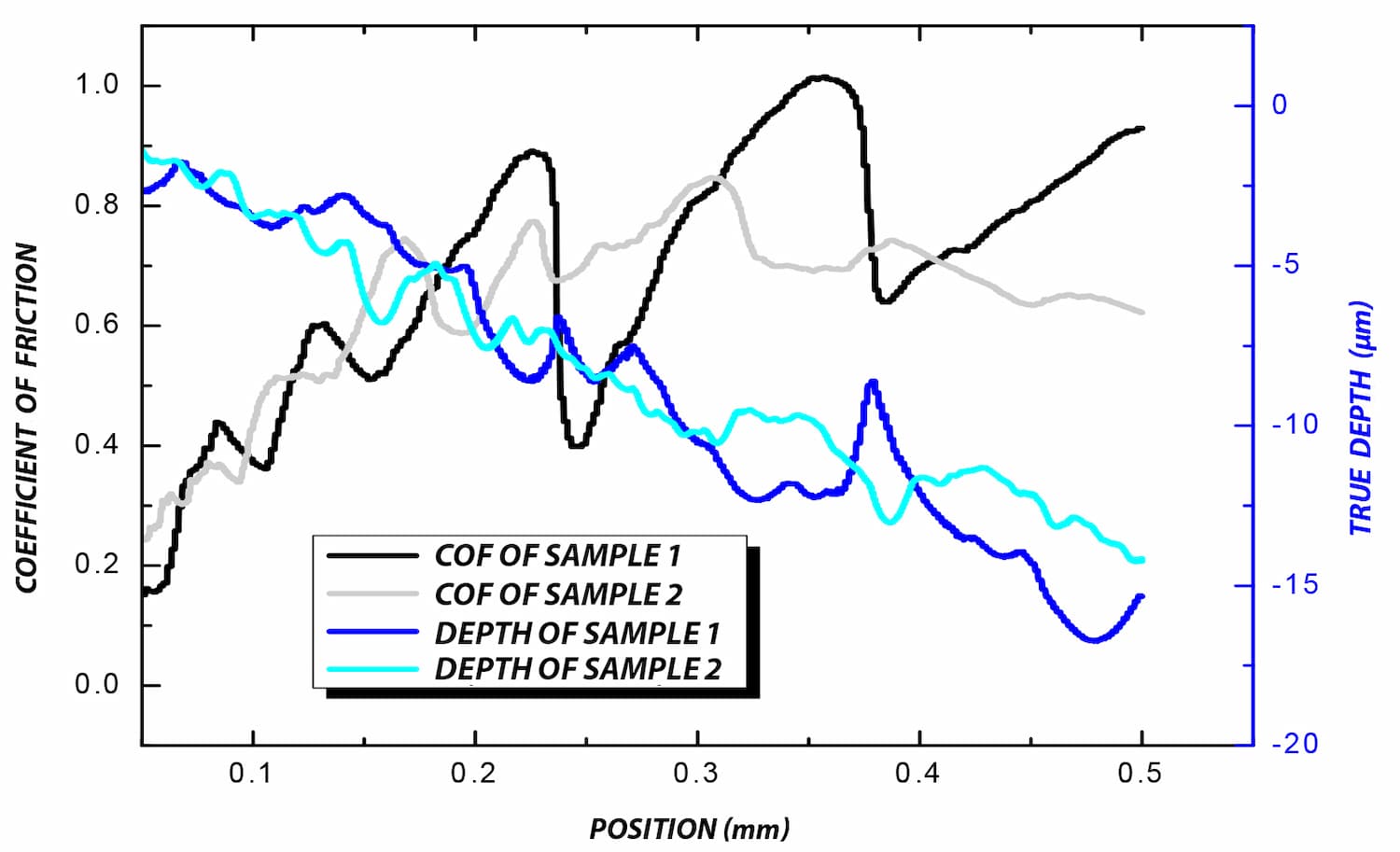
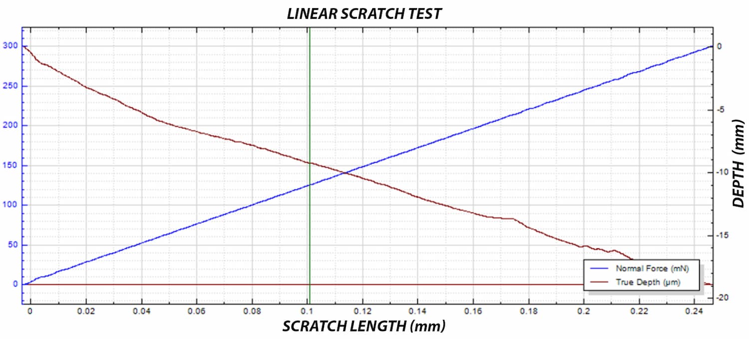
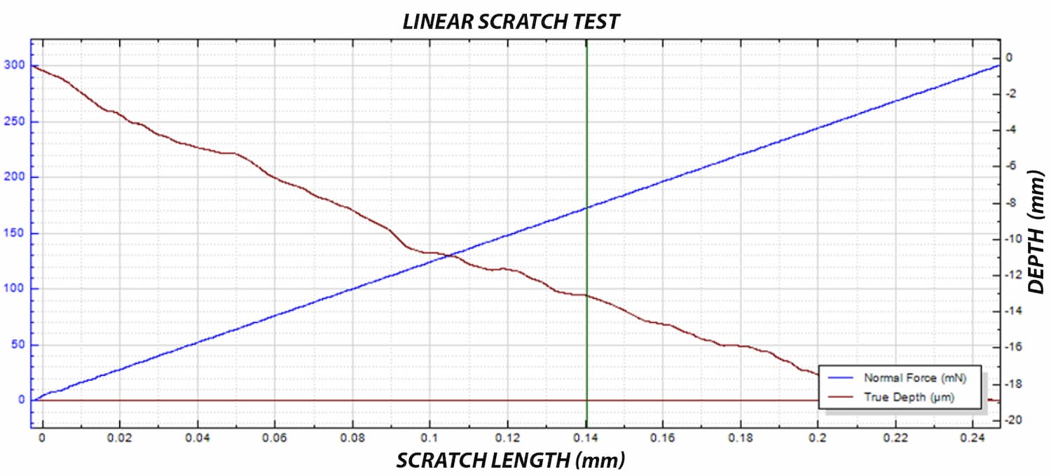
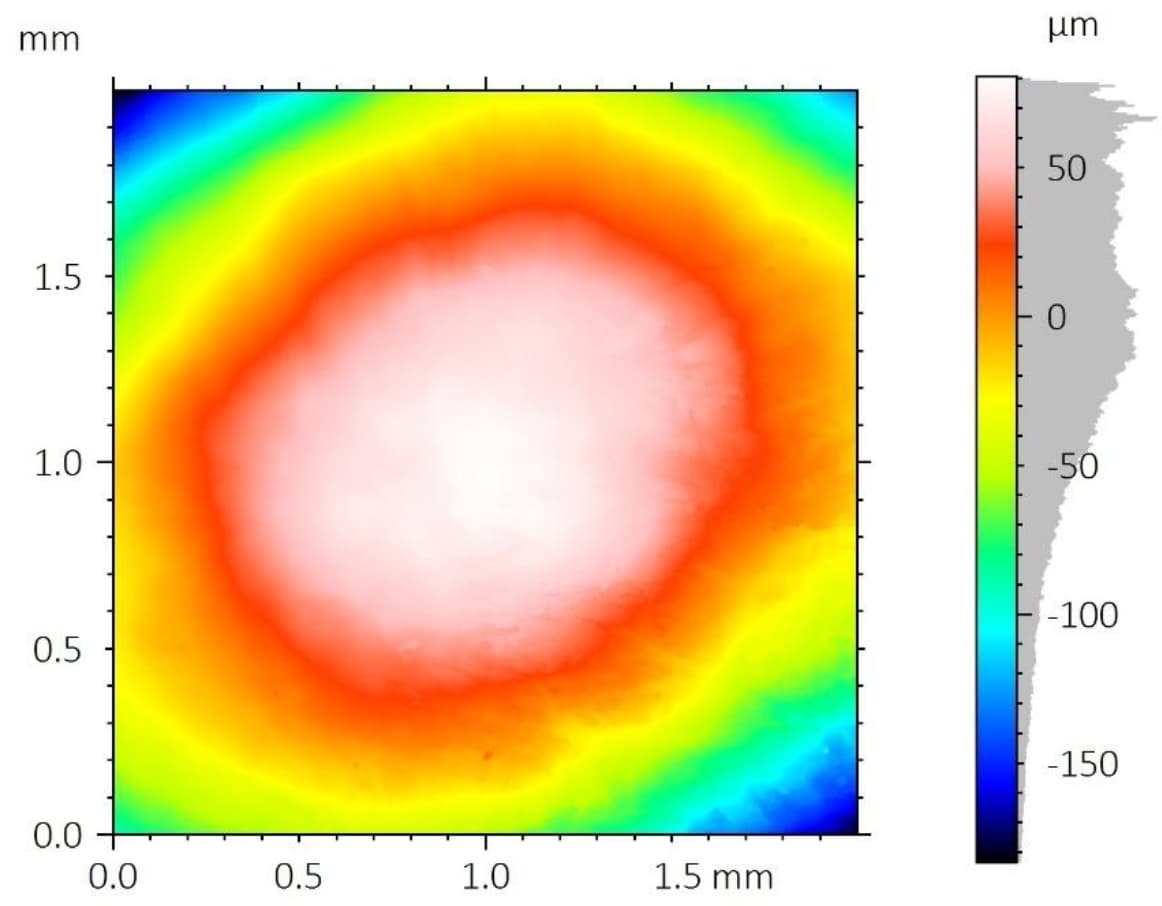
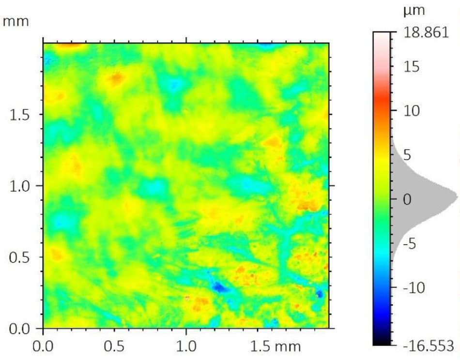
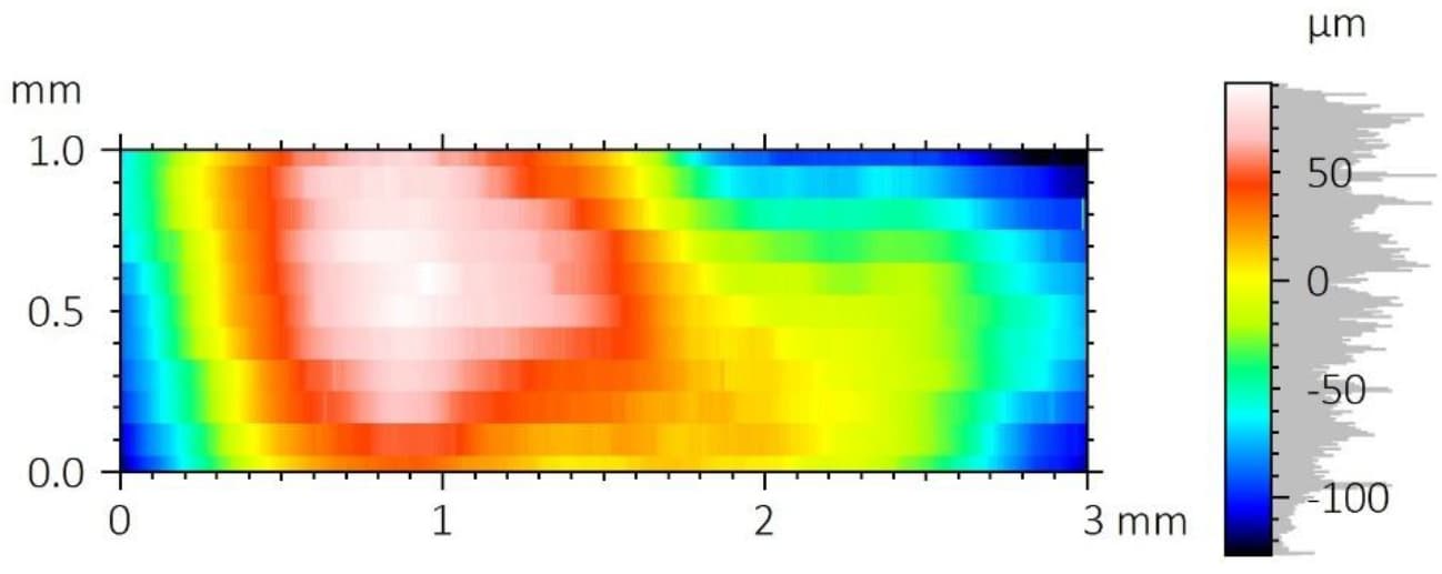
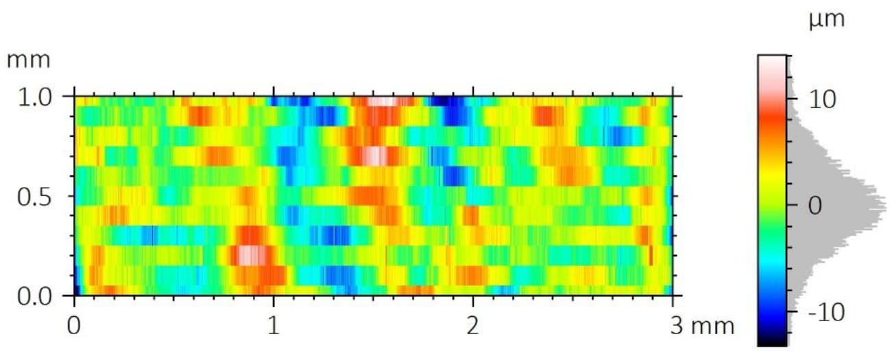
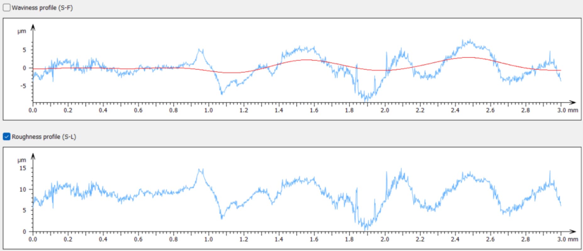
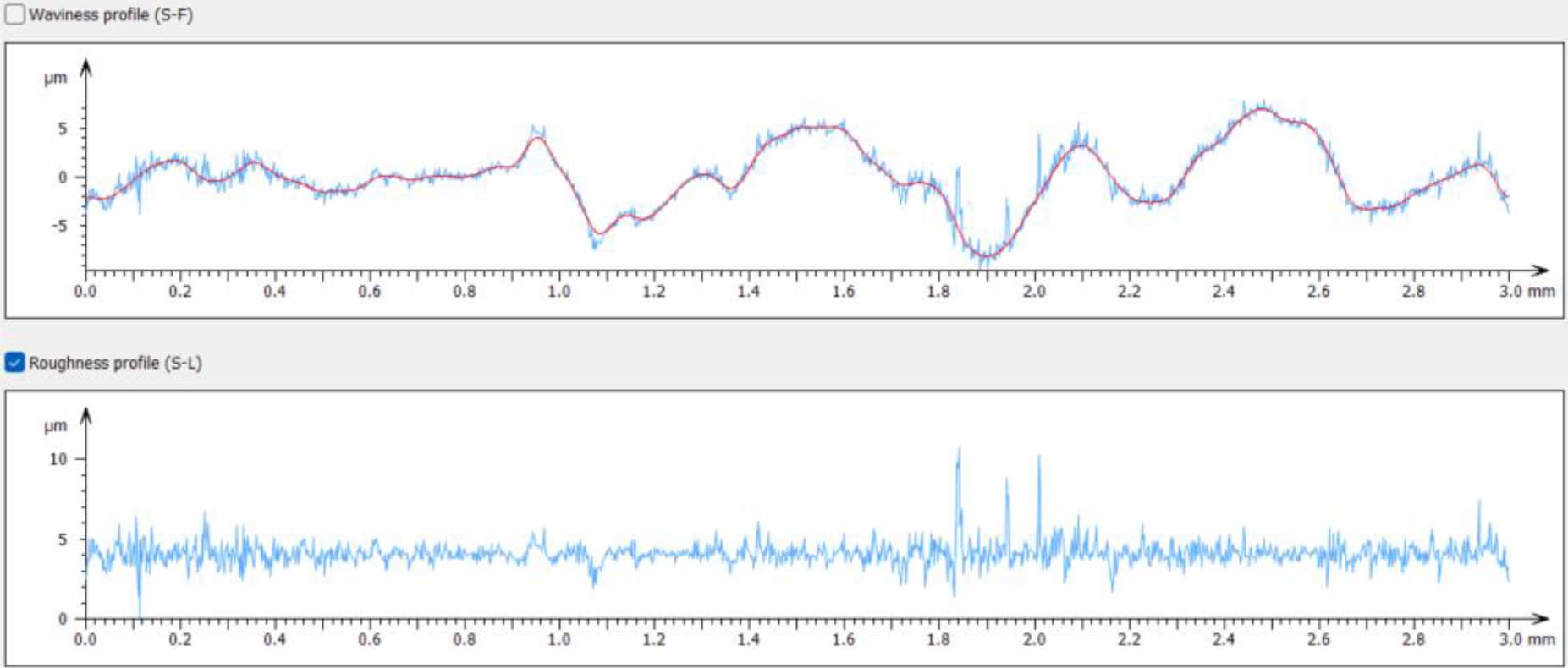
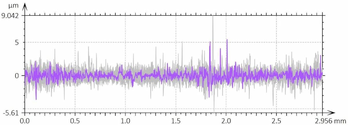
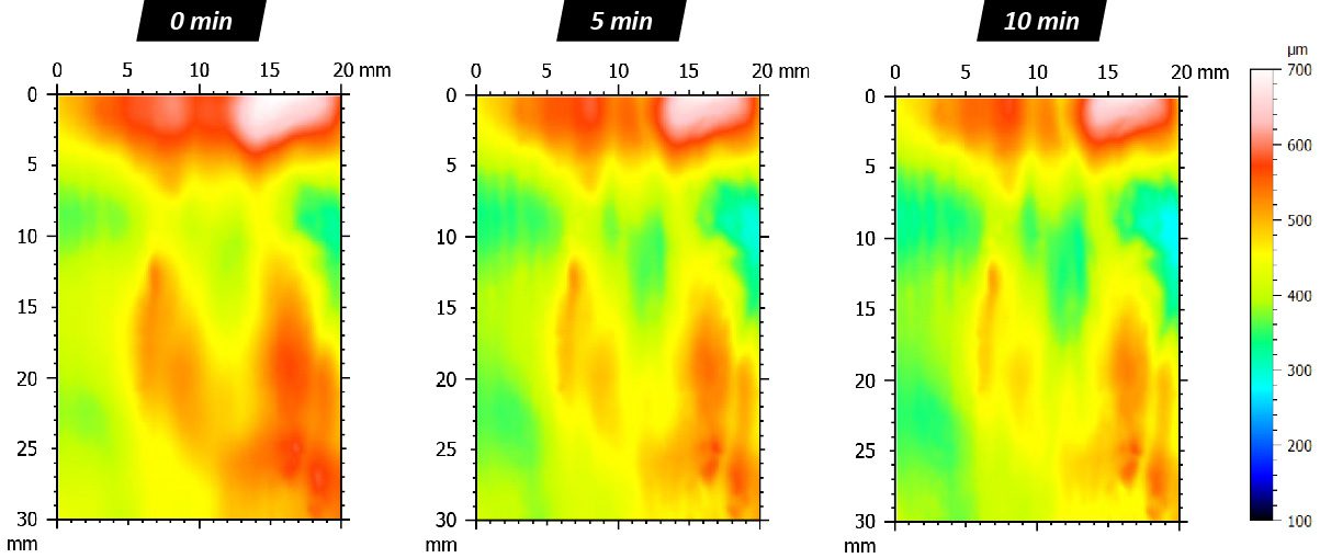
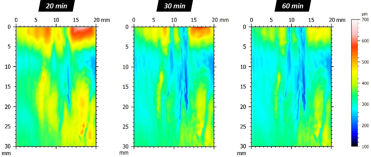
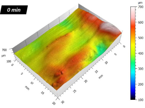
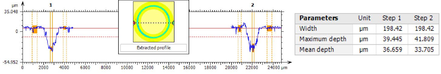
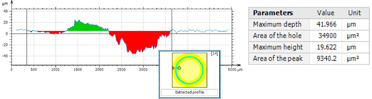
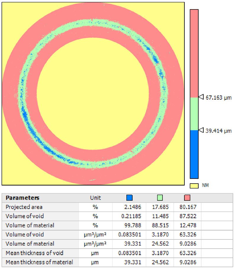
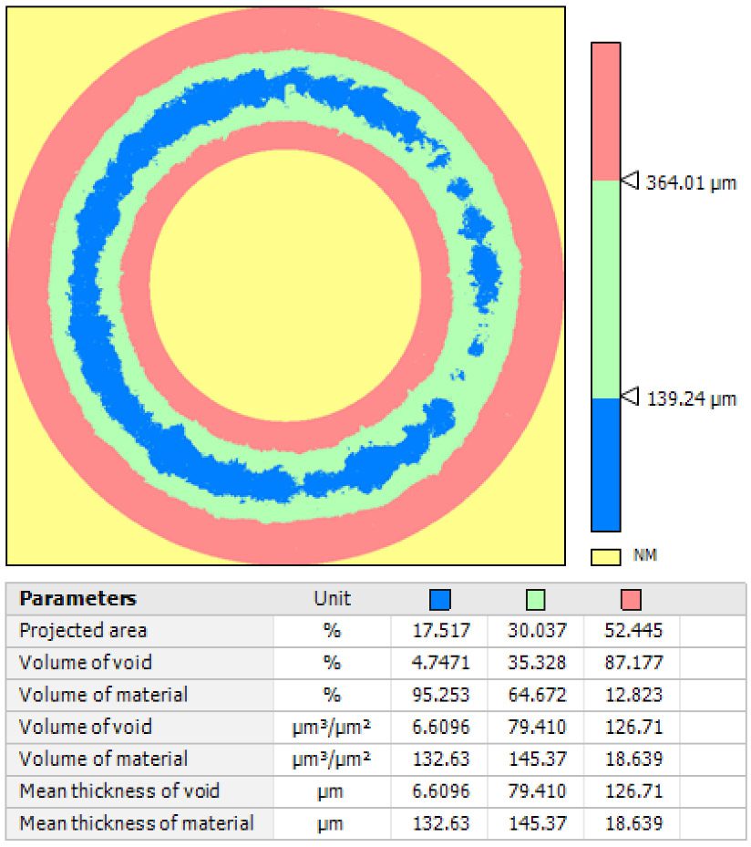
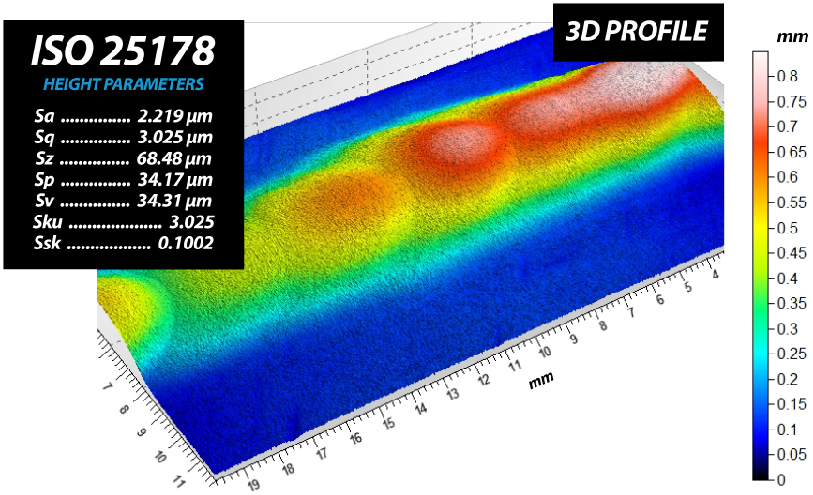
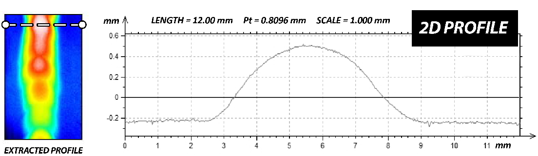
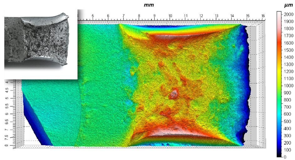
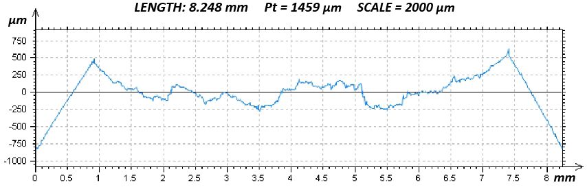
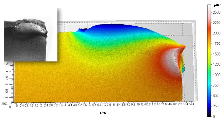
A critical concern in these systems is the delamination of the polymer coating from the metallic stent substrate. This coating carries the drug-eluting layer, and its adhesion directly impacts device performance and reliability.

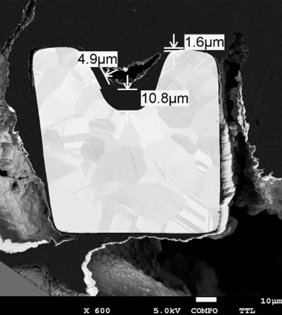
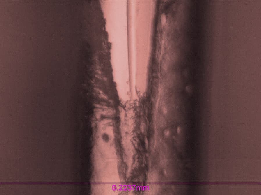
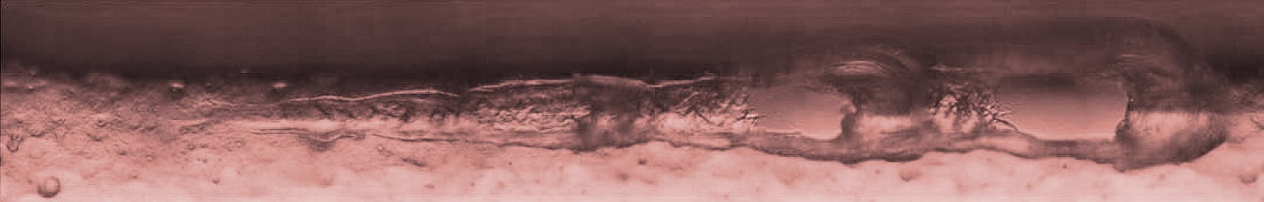
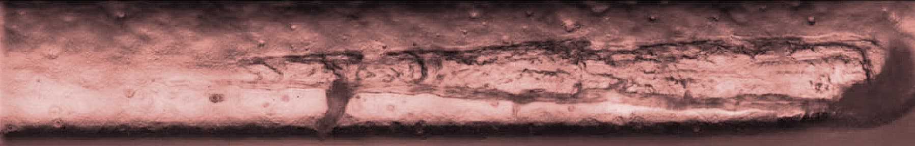
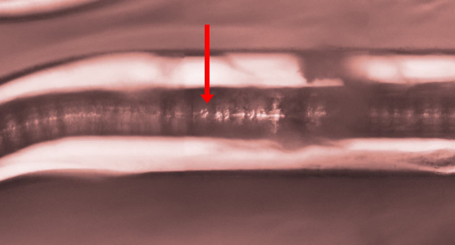
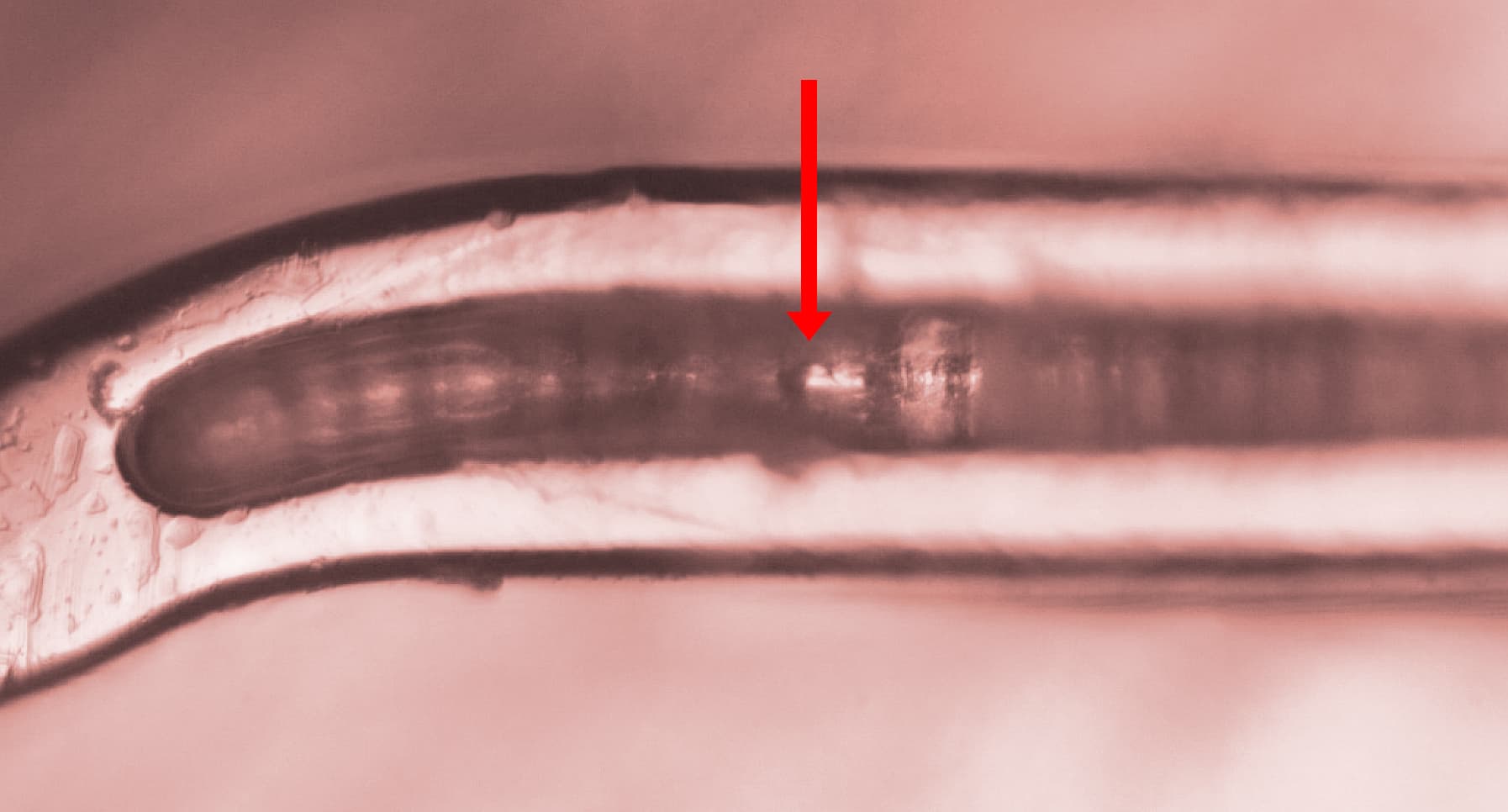
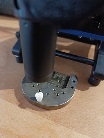
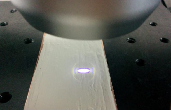
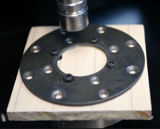
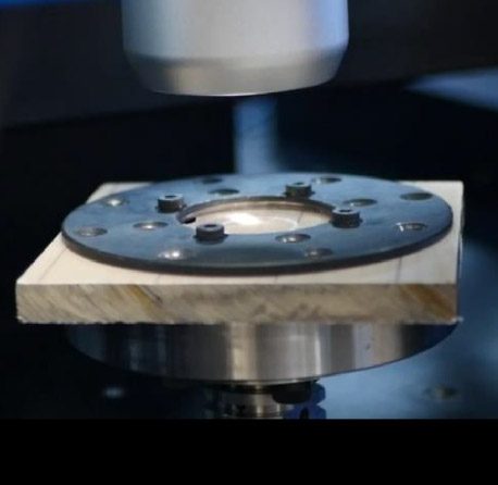
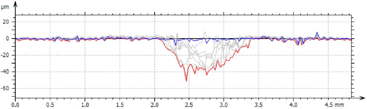
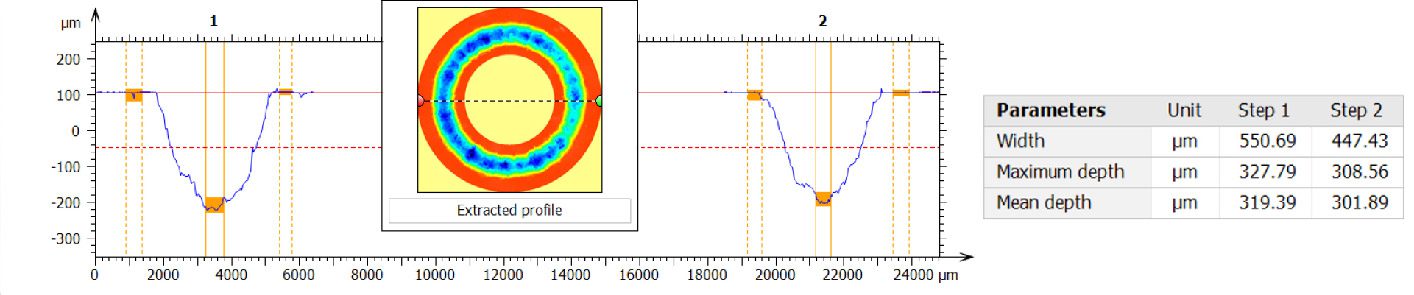
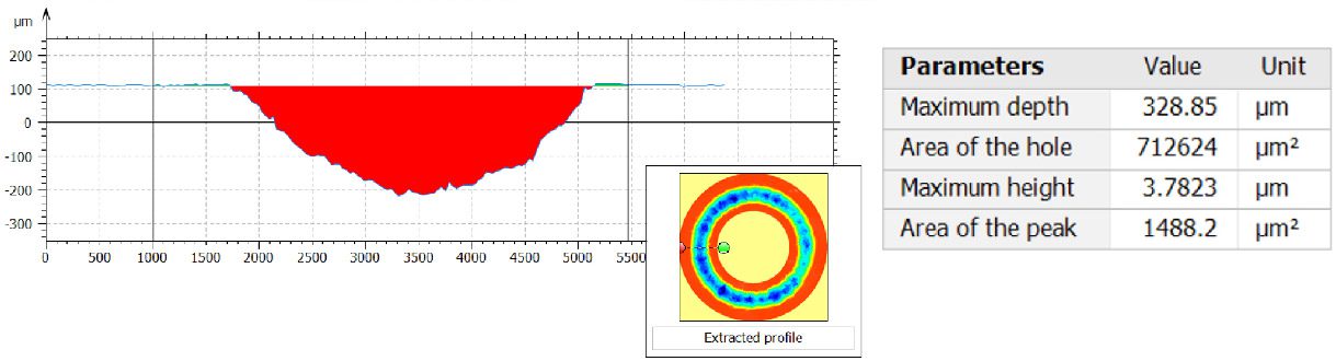
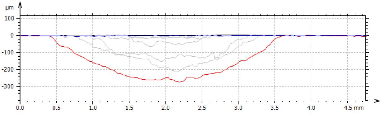
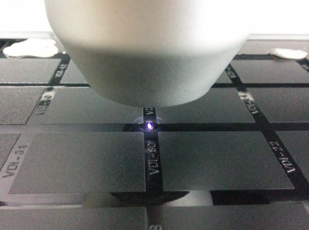
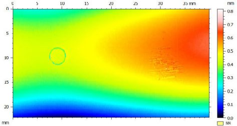
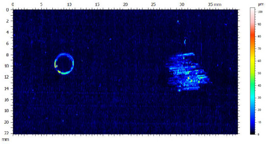
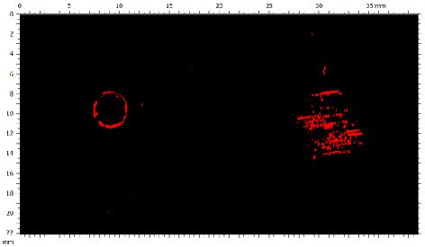
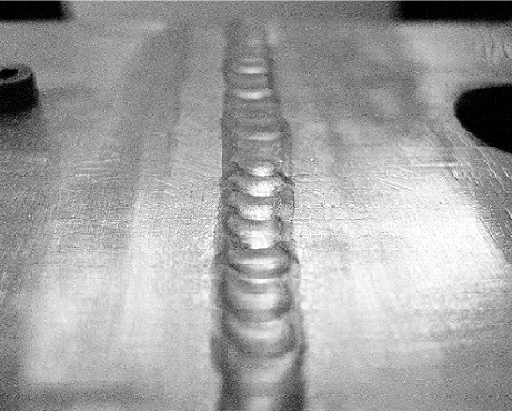
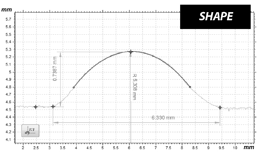
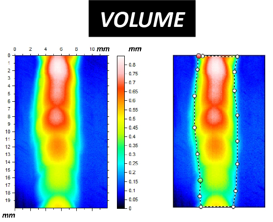
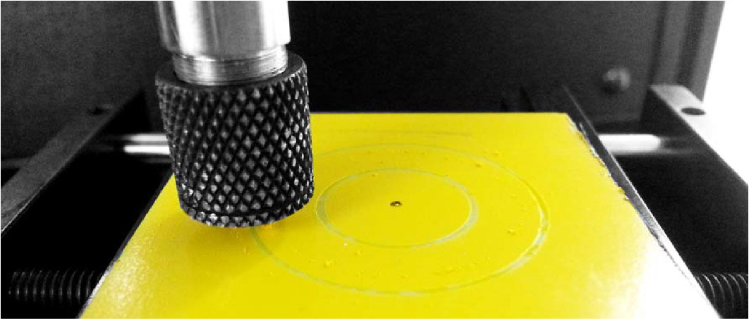
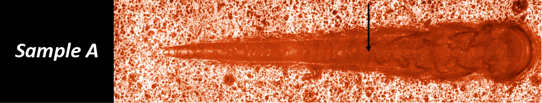
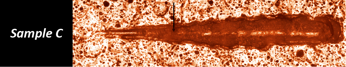
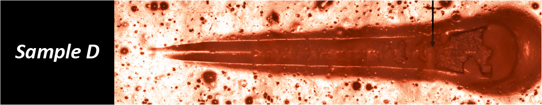
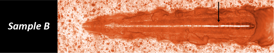
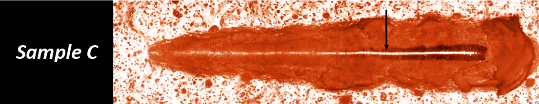
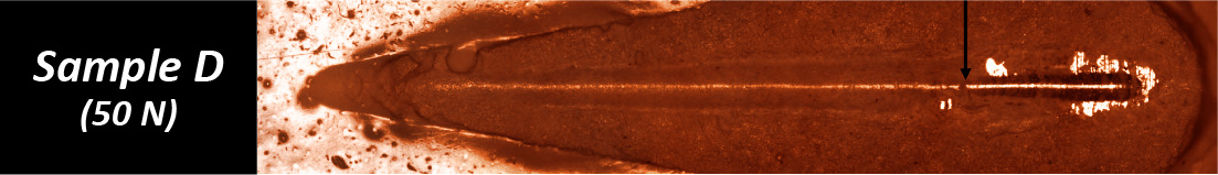
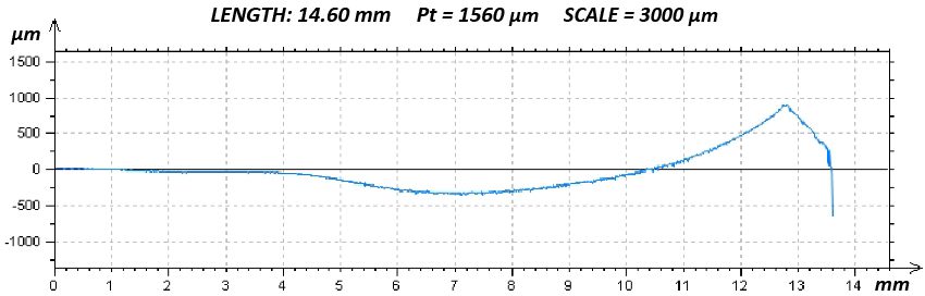
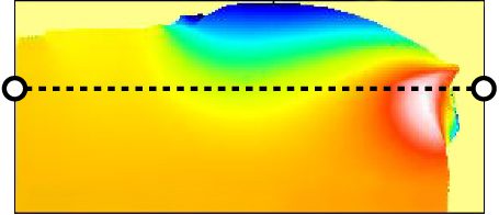
To improve coating adhesion, stents are often designed with complex geometries. In this study, the polymer coating is located at the bottom of grooves within the stent mesh. This configuration presents a significant challenge for adhesion measurement.

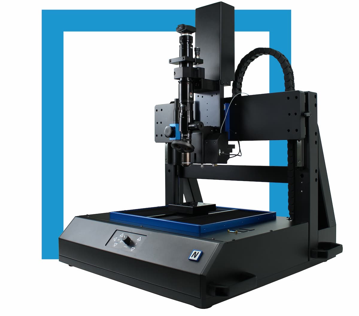
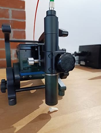
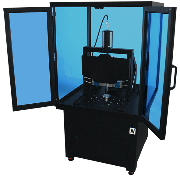
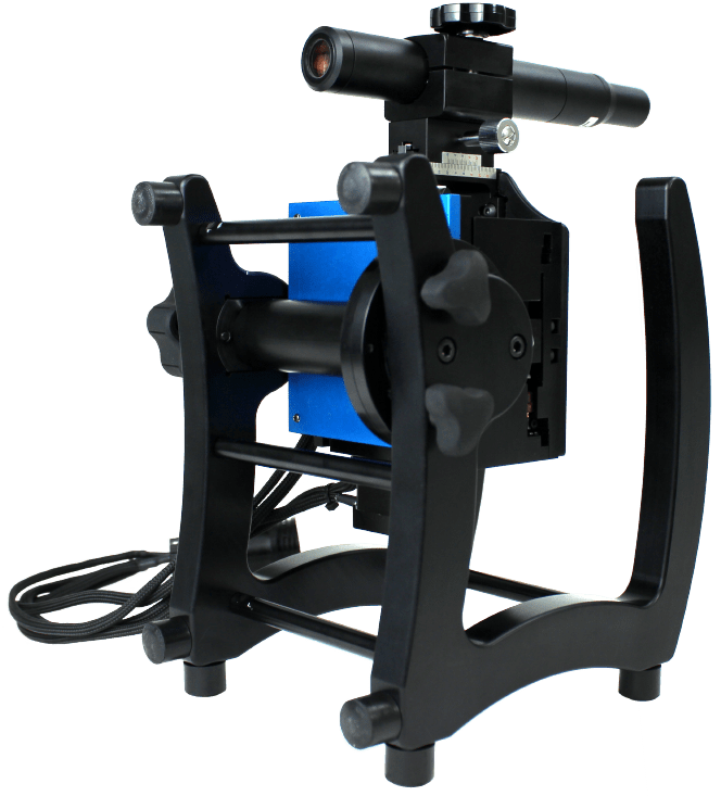
A reliable method is required to quantitatively evaluate the interfacial strength between the polymer coating and the metal substrate. The small diameter of the stent mesh, comparable to a human hair, combined with its three-dimensional geometry, requires:

  • ultrafine X-Y positioning accuracy
  • precise control of applied load
  • accurate depth measurement during testing

Measurement Method

Nano scratch testing is performed using the 나노베아 PB1000 기계식 테스터, in Nano Scratch Mode, to evaluate the cohesive and adhesive strength of the polymer coating on the metal mesh of stent samples.

Controlled scratch measurements are carried out on stent geometries with dimensions comparable to a human hair, enabling precise evaluation of coating adhesion on complex stent structures.

나노비아 PB1000 Advanced

기계 테스터

테스트 조건

1. Regular Stent Samples

The stent is fixed on the sample stage, with a support wire inserted inside the stent tube to ensure stability during nano scratch testing. The NANOVEA Mechanical Tester is used to perform nano scratch measurements using the parameters summarized in Table 1, to evaluate the cohesive and adhesive strength of the polymer coating on the metal substrate.

ParameterValue
Load type프로그레시브
Initial load0.05 mN
Final load300 and 100 mN
Sliding speed0.5 mm/min
Sliding distance0.5 mm
Indenter geometry원뿔형
Indenter material (tip)다이아몬드
들여쓰기 팁 반경20 µm
온도24°C (room)

표 1: Test parameters for nano scratch measurements on regular stent samples

2. Grooved Stent Samples

The SEM image in Fig. 1 shows the cross-section of the stent sample. The stent features a groove with a depth of approximately 30 µm. The polymer coating, with a thickness of 10.8 µm, is located at the bottom of the groove.

Standard 60° conical diamond tips are not sharp enough to reach the bottom of the groove without contacting the sidewalls. Therefore, a sharper 40° conical diamond tip is used in this study (Fig. 2).

Nano scratch measurements are performed using the parameters summarized in Table 2.

Parameter Value
Load type 프로그레시브
Initial load 0.1 mN
Final load 300 mN
로딩 속도 300 mN/min
Scratch length 0.25 mm
Scratch speed 0.25 mm/min
Indenter geometry 40° cone
Indenter material (tip) 다이아몬드
들여쓰기 팁 반경 5 µm

Table 2: Test parameters for nano scratch measurements on grooved stent samples

stent groove cross section polymer coating thickness adhesion analysis nano scratch testing

Fig. 1: SEM cross-section of a grooved stent showing polymer coating located at the bottom of the groove, highlighting the challenge of coating adhesion measurement in recessed geometries.

nano scratch diamond tip 40 degree stent groove coating adhesion testing schematic

Fig. 2: Schematic of a 40° conical diamond tip designed for nano scratch testing inside stent grooves, enabling accurate adhesion measurement without sidewall interference.

결과 및 토론

The stent mesh has a diameter of approximately 100 μm, comparable to a human hair. Precise positioning is therefore critical to ensure the scratch test is performed at the center of the stent mesh. The NANOVEA Mechanical Tester provides X–Y positioning accuracy down to 0.25 μm, enabling accurate test placement under the integrated optical microscope.

1. Regular Stent Samples

Nano scratch testing is performed with a progressively increasing load up to 300 mN. The full scratch track on the stent is shown in Fig. 3a, while failure behavior at different stages is presented in Fig. 3b and 3c.

Two critical loads are identified:

  • Lc1: the load at which the first visible damage appears on the coating
  • Lc2: the load at which the coating is fully removed and the substrate is exposed

The evolution of coefficient of friction (COF) and penetration depth is shown in Fig. 4, providing insight into the progression of coating failure during the test.

The first signs of coating damage appear at Lc1 ≈ 14.5 mN. As the applied load increases, the diamond tip progressively penetrates the polymer coating, resulting in a wider and deeper scratch track. During this phase, the COF increases from approximately 0.05 to 0.7.

At Lc2 ≈ 78.1 mN, the coating is fully delaminated from the metal substrate. Beyond this point, as the load continues to increase, both COF and penetration depth remain relatively stable due to the mechanical support of the underlying metal substrate.

nano scratch track stent coating progressive load adhesion testing

(a) Full Scratch Track

(b) Lc1 ≈ 14.5 mN

stent coating delamination lc2 nano scratch 78.1 mN adhesion testing

(c) Lc2 ≈ 78.1 mN

Fig. 3: Nano scratch track on a stent coating under progressively increasing load, showing (a) full scratch path, (b) initial coating failure at Lc1 ≈ 14.5 mN, and (c) complete coating delamination at Lc2 ≈ 78.1 mN.

nano scratch testing stent coating coefficient of friction depth progression adhesion failure

Fig. 4: Evolution of coefficient of friction (COF) and penetration depth during nano scratch testing of a stent coating under progressively increasing load, showing the progression of coating failure and transition to substrate support.

Failures during nano scratch testing up to a maximum load of 300 mN occur at critical loads below 100 mN. To enable a more quantitative comparison of coating performance, additional tests are performed with a maximum load of 100 mN on two stent samples, referred to as Sample 1 and Sample 2.

Fig. 5 compares the scratch tracks of Sample 1 and Sample 2 after nano scratch testing. Sample 1 exhibits the first sign of coating damage at a critical load of Lc1 ≈ 13.2 mN, while Sample 2 shows initial failure at a higher load of Lc1 ≈ 21.1 mN.

Coating delamination occurs at 62.5 mN for Sample 1. In contrast, the coating on Sample 2 remains intact throughout the test, continuing to protect the metal substrate under the same loading conditions.

This behavior is further reflected in the evolution of coefficient of friction (COF) and penetration depth, as shown in Fig. 6. When the diamond tip penetrates through the coating and contacts the metal substrate in Sample 1, the COF reaches a peak while the penetration depth decreases due to the increased stiffness of the underlying substrate.

stent coating sample 1 early failure nano scratch track delamination adhesion testing

(a) Sample 1 – Early Coating Failure

stent coating sample 2 high adhesion nano scratch track minimal damage testing

(b) Sample 2 – Improved Coating Integrity

Fig. 5: Comparison of nano scratch tracks for two stent coatings, showing (a) early coating failure and delamination in Sample 1, and (b) improved coating integrity in Sample 2 under the same loading conditions.

nano scratch testing stent coating COF depth comparison sample 1 sample 2 adhesion performance

Fig. 6: Comparison of coefficient of friction (COF) and penetration depth for Sample 1 and Sample 2 during nano scratch testing, showing earlier substrate contact and higher friction response in Sample 1, indicating weaker coating adhesion.

2. Grooved Stent Samples

As shown in Fig. 1 and Fig. 7, the grooved stent mesh has a diameter of approximately 90 μm, comparable to a human hair. The groove has a width of ~50 μm and a depth of 30 μm. This geometry presents a significant challenge for nano scratch testing, particularly for evaluating coating adhesion at the bottom of the groove.

Precise positioning is critical to locate the scratch test within the groove. The nano scratch test is performed with a progressively increasing load up to 300 mN. The full scratch tracks of grooved stent Samples 3 and 4 are compared in Fig. 7.

The critical load Lc is defined as the load at which the coating fails and the substrate becomes exposed. The evolution of normal load and penetration depth, shown in Fig. 8, provides further insight into the progression of coating failure during testing.

As the applied load increases, the diamond tip progressively penetrates the polymer coating, resulting in a deeper scratch track. When the critical load Lc is reached, the coating delaminates from the metal substrate.

Sample 3 exhibits coating failure at Lc ≈ 126 mN, while Sample 4 fails at a higher load of Lc ≈ 173 mN. This difference indicates stronger adhesion of the coating in Sample 4.

The measured critical loads enable quantitative comparison of coating adhesion performance. Under the same testing conditions, the coating on Sample 4 demonstrates higher resistance to delamination, making it the better-performing candidate in this study.

stent groove coating failure sample 3 nano scratch 126 mN adhesion testing

(c) Sample 3 – Coating Failure in Groove (Lc ≈ 126 mN)

stent groove coating adhesion sample 4 nano scratch 173 mN minimal failure testing

(d) Sample 4 – Higher Adhesion in Groove (Lc ≈ 173 mN)

Fig. 7: Nano scratch tracks inside stent grooves for Samples 3 and 4, showing (c) coating failure at Lc ≈ 126 mN in Sample 3 and (d) higher adhesion with delayed failure at Lc ≈ 173 mN in Sample 4.

(a) Sample 3 – Earlier Coating Failure (Lc ≈ 126 mN)

(b) Sample 4 – Delayed Failure and Higher Adhesion (Lc ≈ 173 mN)

Fig. 8: Evolution of normal load and penetration depth during nano scratch testing inside stent grooves for Samples 3 and 4, showing earlier coating failure in Sample 3 and delayed failure at higher load in Sample 4. The vertical green line indicates the critical load (Lc) where coating delamination occurs.

결론

This study demonstrates the ability of the NANOVEA Mechanical Tester to quantitatively evaluate the cohesive and adhesive strength of polymer coatings on both regular and grooved stent geometries using nano scratch testing.

The recessed geometry of the stent grooves, approximately 50 μm wide and 30 μm deep, presents a significant challenge for coating adhesion measurement. The high X–Y positioning accuracy of 0.25 μm enables precise placement of the scratch test within these confined regions, allowing direct evaluation of coating performance where failure is most critical.

By applying a controlled, progressively increasing load, critical loads associated with coating failure can be identified and compared across samples. This approach enables reliable differentiation of coating adhesion performance and interfacial integrity, even on small, complex stent structures.

참조

[I] http://www.nhlbi.nih.gov/health/health-topics/topics/stents
[II] http://www.scielo.org.mx/scielo.php?script=sci_arttext&pid=S1405-99402006000300008

Frequently Asked Questions About Stent Coating Adhesion Testing

What is stent coating adhesion testing?

Stent coating adhesion testing evaluates how strongly a polymer coating is bonded to the metal substrate of a stent. Techniques such as nano scratch testing quantify the load at which coating damage and delamination occur, providing measurable indicators of adhesion strength.

What is critical load (Lc) in nano scratch testing?

Critical load (Lc) is the applied load at which a coating fails during a scratch test.

  • Lc1 corresponds to the first visible damage in the coating
  • Lc2 indicates complete coating removal and exposure of the substrate

These values are used to quantify and compare coating adhesion performance.

Why is coating adhesion important in drug-eluting stents?

Coating adhesion directly affects the reliability of drug-eluting stents. Poor adhesion can lead to coating delamination, which may compromise controlled drug release and increase the risk of device failure.

How do you measure coating adhesion inside stent grooves?

Measuring adhesion inside stent grooves requires high positioning accuracy and appropriate indenter geometry. Nano scratch testing with sharp diamond tips allows access to recessed coating regions, enabling direct evaluation of adhesion within complex stent geometries.

What does coefficient of friction (COF) indicate in scratch testing?

The coefficient of friction (COF) reflects changes in surface interaction during the scratch test. A sudden increase in COF often indicates coating failure and contact between the indenter and the underlying metal substrate.

How can nano scratch testing compare different coating formulations?

Nano scratch testing enables direct comparison of coatings by measuring critical loads under controlled conditions. Higher critical loads indicate stronger adhesion and improved resistance to delamination, allowing selection of better-performing coating systems.

Dentist holding dental model for tooth surface roughness analysis and 3D reconstruction

Dental Surface Roughness Measurement & 3D Tooth Topography

Application Note | Dental Surface Characterization

Dental Surface Roughness Measurement and Full 3D Tooth Topography

Surface Roughness Analysis Using Non-Contact Optical Profilometry

Dental surface roughness measurement and 3D molar reconstruction using optical profilometry

작성자

Walter Alabiso, PhD; Davide Morrone, MPhys; Andrew Shore, MA

소개

The ability to accurately characterize tooth surfaces, including micro-roughness and 3D surface topography at the nanometer scale, enables advanced research and applications in orthodontics and dental materials science. Non-contact optical profilometry provides a precise method for measuring dental surface roughness and analyzing tooth surface morphology without damaging delicate structures. These measurements support the development of composite dental materials that replicate the natural surface roughness of enamel, as well as the design and fabrication of patient-specific dental casts and restorative components.

Low surface roughness plays a primary role in limiting bacterial adhesion and plaque formation, thereby reducing the risk of cavities. An increase in average roughness (Ra) above 2 µm leads to a steep increase in biofilm formation in vivo.¹ An Ra of 0.2 µm is considered the threshold value below which no further reduction in bacterial adhesion can be expected.²

Reconstruction of the tooth’s 3D surface topography enables the fabrication of dental casts, which are essential for accurate diagnosis, treatment planning, and the fabrication of dental appliances.

Non-Contact Optical Profilometry for Dental Surface Analysis

The present study illustrates the potential of NANOVEA’s high-precision non-contact optical profilometers for dental surface roughness measurement and 3D tooth topography analysis. Chromatic Light technology offers significant advantages over classical touch probe techniques. It acquires data points from deep crevices and complex geometries without introducing measurement errors or artifacts caused by local plastic deformation and without requiring extensive data manipulation.

Compared to focus variation systems, single-point optical sensing provides superior lateral and height accuracy, with X/Y resolution below 0.5 µm, maximum vertical resolution of 1.9 nm, and the ability to measure surface angles up to 87°. The technique is effective on transparent, opaque, specular, diffusive, polished, and rough dental surfaces, making it well suited for comprehensive dental surface characterization.

Measurement Method

이 애플리케이션에서는 NANOVEA JR25 Non-Contact Optical Profiler was used to analyze the surface roughness and 3D surface topography of an adult human molar previously affected by tooth decay. The side of the tooth was scanned using a PS2–MG140 single-point optical sensor to measure surface roughness parameters over a defined region of interest and along multiple line profiles.

The crown of the tooth was then scanned and reconstructed using a PS5–MG35 single-point optical sensor, which is suited for larger-area acquisition and full 3D tooth topography measurement.


나노비아 JR25 Portable
광학 프로파일로미터

Surface Measurement Using NANOVEA Optical Profilometer

Surface roughness measurements were performed on the lateral side of the molar crown, followed by full 3D reconstruction of the crown surface. Separate single-point optical sensors were used to optimize measurement accuracy for both localized roughness analysis and large-area surface topography acquisition.

PS2 – MG140

Surface roughness analysis by area and parallel line profiles on the side of the tooth’s crown.

PS5 – MG35

Full 3D surface reconstruction of the tooth’s crown.

측정 매개변수

The following measurement parameters were used for localized surface roughness analysis and full 3D surface reconstruction of the molar crown using NANOVEA single-point optical sensors.

ParameterRoughness Analysis (Area)Roughness Analysis (Profiles)Full 3D Reconstruction
Optical PenPS2-MG140PS2-MG140PS5-MG35
Z-Range [µm]30030010000
X-Distance [mm]2.003.007.50
X-Step Size [µm]1.701.7010.00
Y-Distance [mm]2.001.007.00
Y-Step Size [µm]1.70100.0010.00
Average (Avg)111
Measurement TypeDirectDirectDirect
Acquisition ModeSingle FrequencySingle FrequencyDouble Frequency
Acquisition Rate [Hz]200200100–400
Light Intensity [%]100100100

Optical Profilometry Results

Surface Roughness Analysis (Area)

The PS2 single-point optical sensor was used to investigate fine surface features on the side of the tooth. The image below shows a false-color 2D surface map of the scanned region obtained by non-contact optical profilometry.

False-color 2D height map of scanned tooth surface region

A least-squares degree-8 polynomial form removal was applied to isolate the surface roughness component. The roughness filters S-Gaussian 2.5 µm and L-Gaussian 0.8 mm were then applied according to ISO 25178. The resulting filtered surface and corresponding roughness parameters are presented below.

ISO 25178 – Roughness (S-L)
S-filter (λs): Gaussian, 2.5 µm
F: [Workflow] Form removed (LS-poly 8)
L-filter (λc): Gaussian, 0.8 mm
Height Parameters
Sq2.433µm제곱 평균 제곱근 높이
Ssk-0.102 기울기
SKU3.715 첨도
Sp18.861µm최대 피크 높이
Sv16.553µmMaximum pit depth
Sz35.414µm최대 높이
Sa1.888µm산술 평균 높이

The average surface roughness Sa is 1.888 µm, while the peak-to-valley height Sz reaches 35.414 µm.

A 3D surface rendering of the filtered area is shown below for visualization.

3D rendering of ISO 25178 filtered tooth surface roughness

Roughness Analysis (Profiles)

Surface roughness profiles were measured using a series of 11 parallel line scans along the X direction on the side of the tooth. The false-color 2D surface map of the raw scan is shown below.

False-color 2D raw scan of tooth surface for line roughness profiles

The surface form was removed using a least-squares 8-degree polynomial prior to applying the metrological filters, leaving the residual surface shown below.

A statistical analysis of the measured surface roughness profiles reveals the following line roughness parameters.

Overlay of multiple tooth surface roughness profiles for statistical analysis

ISO 4287 – Roughness (S-L)
F: 없음
S-filter (λs): Gaussian, 2.5 µm
L-filter (λc): Gaussian, 0.8 mm
Evaluation length: All λc (3)
Amplitude Parameters – Roughness Profile
  DescriptionMeanStd devMinMax
RpµmMaximum peak height of the roughness profile5.6830.7614.3156.610
RvµmMaximum valley depth of the roughness profile6.2421.0094.7018.438
RzµmMaximum height of roughness profile11.9251.6769.12315.048
RaµmArithmetic mean deviation of the roughness profile2.0630.2971.7102.629
RqµmRoot-mean-square (RMS) deviation of the roughness profile2.5230.3612.0573.175

ISO 4287 – Roughness (S-L)
F: 없음
S-filter (λs): Gaussian, 2.5 µm
L-filter (λc): Gaussian, 0.8 mm
Evaluation length: All λc (3)
Amplitude Parameters – Roughness Profile
Rpµm
Maximum peak height of the roughness profile
Mean5.683
Std dev0.761
Min4.315
Max6.610
Rvµm
Maximum valley depth of the roughness profile
Mean6.242
Std dev1.009
Min4.701
Max8.438
Rzµm
Maximum height of roughness profile
Mean11.925
Std dev1.676
Min9.123
Max15.048
Raµm
Arithmetic mean deviation of the roughness profile
Mean2.063
Std dev0.297
Min1.710
Max2.629
Rqµm
Root-mean-square (RMS) deviation of the roughness profile
Mean2.523
Std dev0.361
Min2.057
Max3.175

The value of Ra is consistent with the Sa value extracted from the surface area measurement.

Different metrological filters can be applied to distinguish between macroscopic waviness and microscopic surface roughness. For example, a coarser filter cut-off, such as the 8 mm cut-off used with the Robust Gaussian order-2 filter, produces a smoother waviness profile (red) that is less sensitive to sharp local variations and follows the original surface profile more loosely.

Comparison of waviness and roughness profiles on tooth surface using coarse filter

Alternatively, a finer cut-off (e.g., 0.08 mm) enables the analysis of micro-roughness by removing the waviness component that follows the original profile at a larger scale, leaving the finer surface roughness features of the tooth visible.

The microroughness analysis obtained using a 0.08 mm L-Gaussian filter is presented below.

Final microroughness profile of tooth surface after filtering

ISO 4287 – Roughness (S-L)
F: 없음
S-filter (λs): Gaussian, 2.5 µm
L-filter (λc): Gaussian, 0.08 mm
Evaluation length: All λc (37)
Amplitude Parameters – Roughness Profile
  DescriptionMeanStd devMinMax
RpµmMaximum peak height of the roughness profile1.5820.1221.3421.748
RvµmMaximum valley depth of the roughness profile1.4660.1191.2541.661
RzµmMaximum height of roughness profile3.0490.1962.8203.409
RaµmArithmetic mean deviation of the roughness profile0.4950.0470.4230.597
RqµmRoot-mean-square (RMS) deviation of the roughness profile0.6430.0560.5620.762

ISO 4287 – Roughness (S-L)
F: 없음
S-filter (λs): Gaussian, 2.5 µm
L-filter (λc): Gaussian, 0.8 mm
Evaluation length: All λc (3)
Amplitude Parameters – Roughness Profile
Rpµm
Maximum peak height of the roughness profile
Mean5.683
Std dev0.761
Min4.315
Max6.610
Rvµm
Maximum valley depth of the roughness profile
Mean6.242
Std dev1.009
Min4.701
Max8.438
Rzµm
Maximum height of roughness profile
Mean11.925
Std dev1.676
Min9.123
Max15.048
Raµm
Arithmetic mean deviation of the roughness profile
Mean2.063
Std dev0.297
Min1.710
Max2.629
Rqµm
Root-mean-square (RMS) deviation of the roughness profile
Mean2.523
Std dev0.361
Min2.057
Max3.175

Full 3D Tooth Surface Topography Reconstruction

The extended Z-scan range of the PS5 optical sensor enables high-fidelity scanning of the entire tooth crown surface. The resulting 3D surface topography is shown below.

False-color surface topography map of full tooth crown measured with optical profilometer

2D VIEW: 2D surface map of the tooth crown measured with optical profilometry

3D surface reconstruction of molar crown from optical profilometer scan

3D VIEW: High-fidelity 3D rendering of the molar crown surface obtained with optical profilometry

결론

In this application, the NANOVEA JR25 Non-Contact Optical Profiler was used to measure the surface roughness and 3D surface topography of an adult human molar.

Both the area scan and the line profile analysis revealed a roughness Rq of approximately 2.5 µm and an Ra of about 1.9–2.0 µm. These values are consistent with results reported in the literature.³ The use of a narrower L-Gaussian filter with an 80 µm cut-off enabled further investigation of micro-roughness, revealing an Rq of 0.643 µm and an Ra of 0.495 µm.

The full 3D surface topography of the molar crown was reconstructed with high fidelity. The high measurement resolution allows detection of fine surface features and crevices. The resulting surface data can be easily processed and exported as STL files, enabling the design and fabrication of customized dental devices and restorative components.

참조

[1] Shin, B.W., et al. Surface Roughness of Prefabricated Pediatric Zirconia Crowns Following Simulated Toothbrushing. Pediatric Dentistry 44.5 (2022): 363–367.
[2] Bollen, C.M.L., Paul Lambrechts, and Marc Quirynen. Comparison of surface roughness of oral hard materials to the threshold surface roughness for bacterial plaque retention: A review of the literature. Dental Materials 13.4 (1997): 258–269.
[3] Suputtamongkol, K., et al. Surface roughness resulting from wear of lithia-disilicate-based posterior crowns. Wear 269.3–4 (2010): 317–322.

Frequently Asked Questions About Dental Surface Roughness Measurement

What is dental surface roughness measurement?

Dental surface roughness measurement quantifies the microscopic texture of tooth surfaces using parameters such as Ra, Rq, and Sa. Optical profilometers measure these features without contacting the surface, allowing accurate analysis of enamel, restorative materials, and dental crowns.

Why use optical profilometry to measure tooth roughness?

Optical profilometry provides non-contact surface measurement with nanometer-scale vertical resolution. It captures 2D surface maps and full 3D surface topography of dental structures without damaging soft or polished surfaces.

What roughness parameters are used for dental surface analysis?

Common roughness parameters include Ra (arithmetic mean roughness), Rq (root mean square roughness), Sa (areal roughness), and Sz (maximum surface height). These parameters help evaluate enamel wear, plaque adhesion risk, and the performance of restorative materials.

Why is surface roughness important in dentistry?

Surface roughness affects plaque retention, wear resistance, and the long-term performance of dental restorations. Controlling micro-roughness can reduce bacterial adhesion and improve the durability of dental materials.

Need Reliable Surface Roughness Measurement for Dental Materials?

Shot Peened 표면 분석

숏핀 표면 분석

3D 비접촉식 프로파일로미터 사용

작성자

크레이그 레싱

소개

쇼트 피닝은 기판에 구형 금속, 유리 또는 세라믹 비드(일반적으로 "샷"이라고 함)를 표면에 가소성을 유도하기 위한 힘으로 충격을 가하는 공정입니다. 피닝 전후의 특성을 분석하면 프로세스 이해 및 제어를 향상시키는 데 중요한 통찰력을 얻을 수 있습니다. 샷에 의해 남겨진 딤플의 표면 거칠기와 적용 범위는 특히 주목할 만한 측면입니다.

숏핀 표면 분석을 위한 3D 비접촉 프로파일로미터의 중요성

전통적으로 샷 피닝된 표면 분석에 사용되었던 기존 접촉식 프로파일로미터와 달리 3D 비접촉식 측정은 완전한 3D 이미지를 제공하여 적용 범위와 표면 지형에 대한 보다 포괄적인 이해를 제공합니다. 3D 기능이 없으면 검사는 2D 정보에만 의존하게 되므로 표면 특성화에 충분하지 않습니다. 지형, 적용 범위 및 거칠기를 3D로 이해하는 것이 피닝 공정을 제어하거나 개선하기 위한 최선의 접근 방식입니다. 나노베아의 3D 비접촉 프로파일로미터 가공된 표면과 피닝된 표면에서 발견되는 가파른 각도를 측정하는 고유한 기능을 갖춘 Chromatic Light 기술을 활용합니다. 또한 프로브 접촉, 표면 변화, 각도 또는 반사율로 인해 다른 기술이 신뢰할 수 있는 데이터를 제공하지 못하는 경우 NANOVEA Profilometer가 성공합니다.

측정 목표

이 응용 분야에서 NANOVEA ST400 비접촉식 프로파일로미터는 비교 검토를 위해 원료와 두 개의 서로 다른 피닝 표면을 측정하는 데 사용됩니다. 3D 표면 스캔 후 자동으로 계산할 수 있는 끝없는 표면 매개변수 목록이 있습니다. 여기에서는 3D 표면을 검토하고 거칠기, 딤플 및 표면적을 정량화하고 조사하는 것을 포함하여 추가 분석을 위해 관심 영역을 선택합니다.

나노비아 ST400 표준
광학 3D 프로파일로미터

샘플

샷 피닝 처리된 표면 시험

결과

강철 표면

샷 피닝 처리된 표면 거칠기
샷 피닝 처리된 표면 특성 분석

ISO 25178 3D 조도 매개변수

SA 0.399㎛ 평균 거칠기
Sq 0.516㎛ RMS 거칠기
Sz 5.686μm 최대 피크-밸리
Sp 2.976μm 최대 피크 높이
Sv 2.711μm 최대 피트 깊이
SKU 3.9344 첨도
Ssk -0.0113 기울기
남자 이름 0.0028mm 자동 상관 길이
Str 0.0613 텍스처 종횡비
스다르 26.539mm² 표면적
Svk 0.589㎛ 감소된 계곡 깊이
 

결과

피닝된 표면 1

샷 피닝 처리된 표면 프로파일
샷 피닝 처리된 표면의 프로파일 측정법

표면 커버리지 98.105%

샷 피닝 처리된 표면 연구

ISO 25178 3D 조도 매개변수

Sa 4.102㎛ 평균 거칠기
Sq 5.153㎛ RMS 거칠기
Sz 44.975μm 최대 피크-밸리
Sp 24.332㎛ 최대 피크 높이
Sv 20.644μm 최대 피트 깊이
SKU 3.0187 첨도
Ssk 0.0625 기울기
남자 이름 0.0976mm 자동 상관 길이
Str 0.9278 텍스처 종횡비
스다르 29.451mm² 표면적
Svk 5.008μm 감소된 계곡 깊이

결과

피닝된 표면 2

샷 피닝 처리된 표면 시험
샷 피닝 처리된 표면의 분석

표면 커버리지 97.366%

샷 피닝 처리된 표면 계측학

ISO 25178 3D 조도 매개변수

Sa 4.330㎛ 평균 거칠기
Sq 5.455㎛ RMS 거칠기
Sz 54.013㎛ 최대 피크-밸리
Sp 25.908μm 최대 피크 높이
Sv 28.105μm 최대 피트 깊이
SKU 3.0642 첨도
Ssk 0.1108 기울기
남자 이름 0.1034mm 자동 상관 길이
Str 0.9733 텍스처 종횡비
스다르 29.623mm² 표면적
Svk 5.167μm 감소된 계곡 깊이

결론

이 숏 피닝 표면 분석 애플리케이션에서 우리는 NANOVEA ST400 3D 비접촉식 프로파일러가 피닝 표면의 지형과 나노미터 세부 사항을 모두 정확하게 특성화하는 방법을 시연했습니다. 표면 1과 표면 2 모두 원료와 비교할 때 여기에 보고된 모든 매개변수에 상당한 영향을 미친다는 것이 분명합니다. 이미지를 간단하게 육안으로 검사하면 표면 간의 차이를 알 수 있습니다. 이는 커버리지 영역과 나열된 매개변수를 관찰하여 추가로 확인됩니다. 표면 2와 비교하여 표면 1은 더 낮은 평균 거칠기(Sa), 더 얕은 찌그러짐(Sv) 및 감소된 표면적(Sdar)을 나타내지만 커버리지 영역은 약간 더 높습니다.

이러한 3D 표면 측정에서 관심 영역을 쉽게 식별하고 거칠기, 마감, 질감, 모양, 지형, 편평도, 뒤틀림, 평면도, 부피, 계단 높이 등을 포함한 포괄적인 측정을 수행할 수 있습니다. 자세한 분석을 위해 2D 단면을 빠르게 선택할 수 있습니다. 이 정보를 통해 표면 측정 리소스의 전체 범위를 활용하여 피닝된 표면을 포괄적으로 조사할 수 있습니다. 통합 AFM 모듈을 사용하여 특정 관심 영역을 추가로 검사할 수 있습니다. NANOVEA 3D 프로파일로미터는 최대 200mm/s의 속도를 제공합니다. 크기, 속도, 스캔 기능 측면에서 사용자 정의할 수 있으며 클래스 1 클린룸 표준도 준수할 수 있습니다. 인라인 또는 온라인 사용을 위한 인덱싱 컨베이어 및 통합과 같은 옵션도 사용할 수 있습니다.

본 문서에 제시된 샘플을 제공해 주신 IMF의 헤이든 씨께 특별히 감사드립니다. 산업 금속 마감 처리 주식회사 | indmetfin.com

페인트 표면 모폴로지

페인트 표면 형태

자동화된 실시간 진화 모니터링
NANOVEA 3D 프로필로미터 사용하기

페인트 표면 모폴로지

작성자

DUANJIE LI, PhD

소개

페인트의 보호 및 장식 특성은 자동차, 해양, 군사 및 건설을 포함한 다양한 산업에서 중요한 역할을 합니다. 내부식성, UV 보호 및 내마모성과 같은 원하는 특성을 달성하기 위해 페인트 공식 및 구조를 신중하게 분석, 수정 및 최적화합니다.

건조 페인트 표면 형태 분석을 위한 3D 비접촉 프로파일로미터의 중요성

페인트는 일반적으로 액체 형태로 적용되며 건조 과정을 거치는데, 여기에는 용매가 증발하고 액체 페인트가 고체 필름으로 변형됩니다. 건조 과정에서 페인트 표면은 모양과 질감이 점진적으로 바뀝니다. 첨가제를 사용하여 페인트의 표면 장력 및 유동 특성을 수정함으로써 다양한 표면 마감 및 질감을 개발할 수 있습니다. 그러나 잘못 공식화된 페인트 레시피 또는 부적절한 표면 처리의 경우 원하지 않는 페인트 표면 손상이 발생할 수 있습니다.

건조 기간 동안 페인트 표면 형태를 정확하게 현장에서 모니터링하면 건조 메커니즘에 대한 직접적인 통찰력을 얻을 수 있습니다. 또한, 표면 형태의 실시간 변화는 3D 프린팅과 같은 다양한 응용 분야에서 매우 유용한 정보입니다. 나노베아 3D 비접촉 프로파일로미터 샘플을 건드리지 않고 재료의 페인트 표면 형태를 측정하여 슬라이딩 스타일러스와 같은 접촉 기술로 인해 발생할 수 있는 모양 변경을 방지합니다.

측정 목표

이 애플리케이션에서는 고속 라인 광학 센서가 장착된 NANOVEA ST500 비접촉 프로파일로미터를 사용하여 1시간 건조 기간 동안 페인트 표면 형태를 모니터링합니다. 우리는 지속적인 형상 변화로 재료의 자동화된 실시간 3D 프로파일 측정을 제공하는 NANOVEA 비접촉 프로파일로미터의 기능을 선보입니다.

나노비아 ST500 대면적
광학 3D 프로파일로미터

결과 및 토론

금속 시트의 표면에 페인트를 도포한 후 고속 라인 센서가 장착된 NANOVEA ST500 비접촉 프로파일로미터를 사용하여 현장에서 즉시 건조 페인트의 형태 변화를 자동 측정했습니다. 0, 5, 10, 20, 30, 40, 50, 60분의 특정 시간 간격으로 3D 표면 형태를 자동으로 측정하고 기록하도록 매크로가 프로그래밍되었습니다. 이 자동 스캔 절차를 통해 사용자는 설정된 절차를 순차적으로 실행하여 스캔 작업을 자동으로 수행할 수 있으므로 수동 테스트 또는 반복 스캔에 비해 노력, 시간 및 발생할 수 있는 사용자 오류를 크게 줄일 수 있습니다. 이 자동화는 서로 다른 시간 간격으로 여러 번 스캔하는 장기 측정에 매우 유용합니다.

광학 라인 센서는 그림 1과 같이 192개의 포인트로 구성된 밝은 라인을 생성합니다. 이 192개의 광점은 샘플 표면을 동시에 스캔하여 스캔 속도를 크게 향상시킵니다. 이렇게 하면 각 3D 스캔이 신속하게 완료되어 각 개별 스캔 중에 상당한 표면 변화를 방지할 수 있습니다.

3D 프로파일로미터를 이용한 도장 코팅 분석

그림 1: 건조 중인 페인트의 표면을 스캔하는 광학 라인 센서.

대표적인 시간에 건조 페인트 토포그래피의 가색 보기, 3D 보기 및 2D 프로파일이 각각 그림 2, 그림 3 및 그림 4에 표시됩니다. 이미지의 가색은 쉽게 식별할 수 없는 기능을 쉽게 감지할 수 있도록 합니다. 서로 다른 색상은 샘플 표면의 서로 다른 영역에서 높이 변화를 나타냅니다. 3D 보기는 사용자가 다양한 각도에서 페인트 표면을 관찰할 수 있는 이상적인 도구를 제공합니다. 테스트의 처음 30분 동안, 페인트 표면의 가색은 따뜻한 톤에서 차가운 톤으로 점차 바뀌어 이 기간 동안 높이가 점진적으로 감소함을 나타냅니다. 30분과 60분에 페인트를 비교할 때 온화한 색상 변화로 알 수 있듯이 이 프로세스가 느려집니다.

페인트 건조 시간의 함수로서 평균 샘플 높이 및 거칠기 Sa 값이 그림 5에 표시되어 있습니다. 0분, 30분 및 60분 건조 시간 후 페인트의 전체 거칠기 분석이 표 1에 나열되어 있습니다. 다음을 관찰할 수 있습니다. 페인트 표면의 평균 높이는 건조 시간의 처음 30분 동안 471에서 329μm로 급격히 감소합니다. 표면 질감은 용매가 증발함과 동시에 발생하여 조도 Sa 값이 7.19에서 22.6μm로 증가합니다. 그 후 페인트 건조 공정이 느려져 샘플 높이와 Sa 값이 60분에 각각 317μm 및 19.6μm로 점진적으로 감소합니다.

이 연구는 건조 페인트의 3D 표면 변화를 실시간으로 모니터링하는 NANOVEA 3D 비접촉 프로파일로미터의 기능을 강조하여 페인트 건조 프로세스에 대한 귀중한 통찰력을 제공합니다. 프로파일로미터는 샘플을 건드리지 않고 표면 형태를 측정함으로써 슬라이딩 스타일러스와 같은 접촉 기술에서 발생할 수 있는 건조되지 않은 페인트의 모양 변경을 방지합니다. 이 비접촉 방식은 건조 페인트 표면 형태에 대한 정확하고 신뢰할 수 있는 분석을 보장합니다.

페인트 표면 모폴로지
도료 코팅 형태학

그림 2: 다른 시간에 건조 페인트 표면 형태의 진화.

도장 표면 특성 분석
도장 표면 프로파일
도장 표면 분석

그림 3: 다양한 건조 시간에 따른 페인트 표면 변화의 3D 보기.

도장 표면 프로파일 측정법

그림 4: 다양한 건조 시간 후 페인트 샘플에 대한 2D 프로파일.

페인트 표면 연구

그림 5: 페인트 건조 시간의 함수로서 평균 샘플 높이 및 거칠기 값 Sa의 변화.

ISO 25178 - 표면 텍스처 매개변수

건조시간(분) 0 5 10 20 30 40 50 60
제곱(µm) 7.91 9.4 10.8 20.9 22.6 20.6 19.9 19.6
SKU 26.3 19.8 14.6 11.9 10.5 9.87 9.83 9.82
SP(µm) 97.4 105 108 116 125 118 114 112
시버트(µm) 127 70.2 116 164 168 138 130 128
사이즈(µm) 224 175 224 280 294 256 244 241
사(µm) 4.4 5.44 6.42 12.2 13.3 12.2 11.9 11.8

제곱 – 제곱 평균 제곱근 높이 | SKU – 첨도 | Sp – 최대 피크 높이 | Sv – 최대 구덩이 높이 | Sz – 최대 높이 | Sv – 산술 평균 높이

표 1: 다른 건조 시간에 페인트 거칠기.

결론

이 애플리케이션에서 우리는 NANOVEA ST500 3D 비접촉 프로파일로미터가 건조 공정 중 페인트 표면 형태의 변화를 모니터링하는 기능을 선보였습니다. 샘플 표면을 동시에 스캔하는 192개의 광점이 있는 라인을 생성하는 고속 광학 라인 센서는 타의 추종을 불허하는 정확도를 보장하면서 연구 시간을 효율적으로 만들었습니다.

획득 소프트웨어의 매크로 기능을 사용하면 3D 표면 형태의 자동 측정을 현장에서 프로그래밍할 수 있으므로 특정 대상 시간 간격에서 여러 스캔을 포함하는 장기 측정에 특히 유용합니다. 시간, 노력 및 사용자 오류 가능성을 크게 줄입니다. 표면 형태의 점진적인 변화는 페인트가 건조됨에 따라 실시간으로 지속적으로 모니터링되고 기록되어 페인트 건조 메커니즘에 대한 귀중한 통찰력을 제공합니다.

여기에 표시된 데이터는 분석 소프트웨어에서 사용할 수 있는 계산의 일부만을 나타냅니다. NANOVEA 프로파일로미터는 투명, 어둡거나 반사 또는 불투명 여부에 관계없이 거의 모든 표면을 측정할 수 있습니다.

트라이보미터를 사용한 바닥재의 점진적 마모 매핑

바닥재 마모 시험

통합 프로파일로미터를 장착한 트라이보미터를 이용한 바닥재의 점진적 마모 매핑

마루 바닥재 마모 시험

작성자

프랭크 리우

소개

바닥재는 내구성이 뛰어나도록 설계되었지만 이동, 가구 사용 등 일상 활동으로 인해 마모되거나 찢어지는 경우가 많습니다. 수명을 보장하기 위해 대부분의 바닥재에는 손상을 방지하는 보호 마모층이 있습니다. 그러나 바닥재 종류와 보행량에 따라 마모층의 두께와 내구성이 달라집니다. 또한 UV 코팅, 장식 층, 유약 등 바닥 구조 내의 다양한 층은 마모율이 다릅니다. 이것이 바로 점진적인 마모 매핑이 필요한 곳입니다. 통합된 NANOVEA T2000 마찰계를 사용하여 3D 비접촉 프로파일로미터, 바닥재의 성능과 수명에 대한 정밀한 모니터링과 분석이 가능합니다. 다양한 바닥재의 마모 거동에 대한 자세한 통찰력을 제공함으로써 과학자와 기술 전문가는 새로운 바닥재 시스템을 선택하고 설계할 때 더 많은 정보를 바탕으로 결정을 내릴 수 있습니다.

바닥 패널에 대한 프로그레시브 마모 매핑의 중요성

바닥재 테스트는 전통적으로 마모에 대한 내구성을 결정하기 위해 샘플의 마모율에 중점을 두었습니다. 그러나 프로그레시브 마모 매핑을 사용하면 테스트 전반에 걸쳐 샘플의 마모율을 분석하여 마모 거동에 대한 귀중한 통찰력을 얻을 수 있습니다. 이러한 심층 분석을 통해 마찰 데이터와 마모율 간의 상관관계를 파악하여 마모의 근본 원인을 파악할 수 있습니다. 마모율은 마모 테스트 전반에 걸쳐 일정하지 않다는 점에 유의해야 합니다. 따라서 마모 진행을 관찰하면 샘플의 마모를 보다 정확하게 평가할 수 있습니다. 기존의 테스트 방법을 뛰어넘는 프로그레시브 마모 매핑의 도입은 바닥재 테스트 분야에서 상당한 발전에 기여했습니다.

3D 비접촉 프로파일로미터가 통합된 NANOVEA T2000 마찰계는 마모 테스트 및 체적 손실 측정을 위한 획기적인 솔루션입니다. 핀과 프로파일로미터 사이를 정밀하게 이동할 수 있는 능력은 마모 트랙 반경이나 위치의 편차를 제거하여 결과의 신뢰성을 보장합니다. 하지만 그게 전부는 아닙니다. 3D 비접촉 프로파일로미터의 고급 기능을 사용하면 고속 표면 측정이 가능해 스캔 시간이 단 몇 초로 단축됩니다. 최대 2,000N의 하중을 적용하고 최대 5,000rpm의 회전 속도를 달성할 수 있는 NANOVEA T2000 트라이보미터 평가 과정에서 다양성과 정확성을 제공합니다. 이 장비가 점진적인 마모 매핑에서 중요한 역할을 한다는 것은 분명합니다.

 
마모계(tribometer)를 이용한 바닥재 마모 시험
프로파일로미터를 이용한 바닥재 마모 시험

그림 1: 마모 테스트 전 샘플 셋업 (왼쪽) 및 마모 테스트 후 마모 트랙의 프로파일 측정(오른쪽).

측정 목표

프로그레시브 마모 매핑 테스트는 석재와 목재 두 가지 유형의 바닥재에 대해 수행되었습니다. 각 샘플은 2, 4, 8, 20, 40, 60, 120초로 테스트 시간을 늘려가며 총 7번의 테스트 주기를 거쳤으며, 시간 경과에 따른 마모를 비교할 수 있도록 했습니다. 각 테스트 사이클이 끝난 후 나노베아 3D 비접촉 프로파일로미터를 사용하여 마모 트랙을 프로파일링했습니다. 프로파일러가 수집한 데이터에서 구멍의 부피와 마모율은 나노베아 트라이보미터 소프트웨어 또는 표면 분석 소프트웨어인 마운틴의 통합 기능을 사용하여 분석할 수 있습니다.

나노비아 T2000 고부하
공압식 마찰계

샘플

마모 매핑 테스트 샘플 목재 및 석재

마모 매핑 테스트 매개변수

로드40 N
테스트 기간다양
속도200 rpm
RADIUS10 mm
거리다양
볼 소재텅스텐 카바이드
볼 지름10 mm

7주기 동안 사용된 테스트 기간은 다음과 같습니다. 2, 4, 8, 20, 40, 60, 120초로 각각 이동했습니다. 이동 거리는 다음과 같습니다. 0.40, 0.81, 1.66, 4.16, 8.36, 12.55, 25.11미터.

마모 매핑 결과

목재 바닥재

테스트 주기최대 COF최소 COFAvg. COF
10.3350.1240.275
20.3370.2070.295
30.3800.2290.329
40.3930.2650.354
50.3520.2050.314
60.3450.1990.312
70.3150.2110.293


방사형 방향

테스트 주기총 부피 손실(µm3총 거리
이동 거리(m)
마모율
(mm/Nm) x10-5
순간 마모율
(mm/Nm) x10-5
12962476870.401833.7461833.746
23552452271.221093.260181.5637
35963713262.88898.242363.1791
48837477677.04530.629172.5496
5120717995115.40360.88996.69074
6147274531827.95293.32952.89311
7185131921053.06184.34337.69599
목재 프로그레시브 마모율 대 총 거리
목재 바닥 마모율

그림 2: 마모율 대비 총 이동 거리(왼쪽)
목재 바닥재의 순간 마모율과 테스트 주기(오른쪽)를 비교한 결과입니다.

마찰 계수 시험
목재 바닥의 점진적 마모 매핑

그림 3: 목재 바닥에서 테스트한 #7의 마모 트랙의 COF 그래프 및 3D 보기.

웨어 매핑 추출 프로파일
바닥재 마모 시험 결과
바닥 표면 특성 분석

그림 4: 테스트 #7의 목재 마모 트랙 단면 분석

프로그레시브 마모 매핑 볼륨 및 면적 분석

그림 5: 목재 샘플 테스트 #7에서 마모 트랙의 부피 및 면적 분석.

마모 매핑 결과

석재 바닥재

테스트 주기최대 COF최소 COFAvg. COF
10.2490.0350.186
20.3490.1970.275
30.2940.1540.221
40.5030.1240.273
50.5480.1060.390
60.5100.1290.434
70.5270.1810.472


방사형 방향

테스트 주기총 부피 손실(µm3총 거리
이동 거리(m)
마모율
(mm/Nm) x10-5
순간 마모율
(mm/Nm) x10-5
1962788460.40595.957595.9573
28042897311.222475.1852178.889
313161478552.881982.355770.9501
431365302157.041883.2691093.013
51082173218015.403235.1802297.508
62017496034327.954018.2821862.899
74251206342053.064233.0812224.187
석재 바닥재 마모율 대 거리
석재 바닥재 순간 마모율 차트

그림 6: 마모율 대비 총 이동 거리(왼쪽)
석재 바닥재의 순간 마모율과 테스트 주기(오른쪽)를 비교합니다.

마루재 마모 마찰학적 시험
마모 트랙의 석재 바닥 3D 프로파일

그림 7: 석재 바닥에서 테스트한 #7의 마모 트랙의 COF 그래프 및 3D 보기.

석재 바닥 프로그레시브 마모 매핑 추출 프로파일
석재 바닥재 추출 프로파일 구멍과 피크의 최대 깊이 및 높이 면적
바닥재 마찰학 시험

그림 8: 테스트 #7의 스톤 마모 트랙 단면 분석.

목재 바닥 프로그레시브 마모 매핑 볼륨 분석

그림 9: 석재 샘플 테스트 #7에서 마모 트랙의 부피 및 면적 분석.

토론

순간 마모율은 다음 공식을 사용하여 계산합니다:
바닥재 공식의 점진적 마모 매핑

여기서 V는 구멍의 부피, N은 하중, X는 총 거리이며, 이 방정식은 테스트 주기 사이의 마모율을 설명합니다. 순간 마모율은 테스트 전반에 걸친 마모율의 변화를 더 잘 파악하는 데 사용할 수 있습니다.

두 샘플의 마모 거동은 매우 다릅니다. 시간이 지남에 따라 목재 바닥재는 높은 마모율로 시작하지만 빠르게 더 작고 일정한 값으로 떨어집니다. 석재 바닥재의 경우 마모율은 낮은 값에서 시작하여 주기에 따라 더 높은 값으로 증가하는 경향을 보입니다. 순간 마모율도 일관성이 거의 없습니다. 이 차이의 구체적인 이유는 확실하지 않지만 샘플의 구조 때문일 수 있습니다. 석재 바닥재는 나뭇결 같은 입자로 이루어져 있어 목재의 촘촘한 구조와 다르게 마모되는 것으로 보입니다. 이러한 마모 현상의 원인을 확인하려면 추가적인 테스트와 연구가 필요합니다.

마찰 계수(COF)의 데이터는 관찰된 마모 거동과 일치하는 것으로 보입니다. 목재 바닥재의 COF 그래프는 사이클 전체에 걸쳐 일관되게 나타나며 꾸준한 마모율을 보완합니다. 석재 바닥재의 경우 사이클에 따라 마모율도 증가하는 것과 유사하게 사이클 전반에 걸쳐 평균 COF가 증가합니다. 마찰 그래프의 모양에도 뚜렷한 변화가 있어 공이 석재 샘플과 상호 작용하는 방식에 변화가 있음을 알 수 있습니다. 이는 사이클 2와 사이클 4에서 가장 두드러집니다.

결론

나노베아 T2000 트라이보미터는 두 개의 서로 다른 바닥재 샘플 사이의 마모율을 분석하여 점진적 마모 매핑을 수행할 수 있는 기능을 선보입니다. 연속 마모 테스트를 일시 중지하고 나노베아 3D 비접촉 프로파일로미터로 표면을 스캔하면 시간에 따른 재료의 마모 거동에 대한 귀중한 통찰력을 얻을 수 있습니다.

3D 비접촉식 프로파일로미터가 통합된 나노베아 T2000 트라이보미터는 COF(마찰 계수) 데이터, 표면 측정, 깊이 판독, 표면 시각화, 체적 손실, 마모율 등을 포함한 다양한 데이터를 제공합니다. 이 포괄적인 정보 세트를 통해 사용자는 시스템과 시료 간의 상호 작용에 대해 더 깊이 이해할 수 있습니다. 제어된 하중, 고정밀, 사용 편의성, 높은 하중, 넓은 속도 범위 및 추가 환경 모듈을 갖춘 NANOVEA T2000 트라이보미터는 마찰학을 한 차원 더 높은 수준으로 끌어올립니다.

3D 프로파일 측정을 이용한 거칠기 매핑 검사

거칠기 매핑 검사

3D 프로파일 측정 사용

작성자

DUANJIE, 박사

소개

표면 거칠기와 질감은 제품의 최종 품질과 성능에 영향을 미치는 중요한 요소입니다. 최상의 처리 및 제어 방법을 선택하려면 표면 거칠기, 질감, 일관성에 대한 철저한 이해가 필수적입니다. 결함이 있는 제품을 적시에 식별하고 생산 라인 조건을 최적화하려면 제품 표면에 대한 빠르고 정량화 가능하며 신뢰할 수 있는 인라인 검사가 필요합니다.

인라인 표면 검사를 위한 3D 비접촉식 프로파일로미터의 중요성

제품의 표면 결함은 재료 가공 및 제품 제조 과정에서 발생합니다. 인라인 표면 품질 검사는 최종 제품의 가장 엄격한 품질 관리를 보장합니다. 나노베아 3D 비접촉식 광학 프로파일러 접촉 없이 샘플의 거칠기를 결정하는 고유한 기능을 갖춘 색채광 기술을 활용합니다. 라인 센서를 사용하면 넓은 표면의 3D 프로파일을 고속으로 스캐닝할 수 있습니다. 분석 소프트웨어에 의해 실시간으로 계산된 거칠기 임계값은 빠르고 안정적인 합격/불합격 도구 역할을 합니다.

측정 목표

이 연구에서는 고속 센서가 장착된 나노베아 ST400을 사용하여 결함이 있는 테프론 샘플의 표면을 검사하여 나노베아의 성능을 보여줍니다.

생산 라인에서 빠르고 안정적인 표면 검사를 제공하는 비접촉식 프로로미터입니다.

나노비아

ST400

결과 및 토론

3D 표면 분석 거칠기 표준 샘플

거칠기 표준의 표면은 그림 1과 같이 192개의 밝은 선을 생성하는 고속 센서가 장착된 나노베아 ST400을 사용하여 스캔했습니다. 이 192개의 포인트가 동시에 샘플 표면을 스캔하기 때문에 스캔 속도가 크게 향상되었습니다.

그림 2는 거칠기 표준 샘플의 표면 높이 맵과 거칠기 분포 맵의 가색 보기를 보여줍니다. 그림 2a에서 거칠기 표준은 각 표준 거칠기 블록의 다양한 색상 그라데이션으로 표시된 것처럼 약간 기울어진 표면을 나타냅니다. 그림 2b에서는 서로 다른 거칠기 블록에서 균일한 거칠기 분포가 나타나며, 색상은 블록의 거칠기를 나타냅니다.

그림 3은 다양한 거칠기 임계값에 따라 분석 소프트웨어에서 생성된 합격/불합격 맵의 예를 보여줍니다. 표면 거칠기가 특정 임계값을 초과하면 거칠기 블록이 빨간색으로 강조 표시됩니다. 이를 통해 사용자는 거칠기 임계값을 설정하여 샘플 표면 마감의 품질을 결정할 수 있습니다.

그림 1: 거칠기 표준 샘플의 광학 라인 센서 스캔

a. 표면 높이 맵:

b. 러프니스 맵:

그림 2: 거칠기 표준 샘플의 표면 높이 맵 및 거칠기 분포 맵의 가색 보기입니다.

그림 3: 거칠기 임계값에 따른 합격/불합격 맵입니다.

결함이 있는 테온 샘플의 표면 검사

테온 샘플 표면의 표면 높이 맵, 거칠기 분포 맵 및 합격/불합격 거칠기 임계값 맵은 그림 4에 나와 있습니다. 표면 높이 맵에 표시된 것처럼 테온 샘플은 샘플의 오른쪽 중앙에 능선 형태가 있습니다.

a. 표면 높이 맵:

그림 4b의 팔레트에서 서로 다른 색상은 로컬 표면의 거칠기 값을 나타냅니다. 거칠기 맵은 테온 샘플의 온전한 영역에서 균일한 거칠기를 나타냅니다. 그러나 움푹 들어간 링과 마모 흉터 형태의 결함은 밝은 색상으로 강조 표시됩니다. 사용자는 그림 4c와 같이 표면 결함을 찾기 위해 합격/불합격 거칠기 임계값을 쉽게 설정할 수 있습니다. 이러한 툴을 통해 사용자는 생산 라인에서 제품 표면 품질을 현장에서 모니터링하고 결함이 있는 제품을 적시에 발견할 수 있습니다. 제품이 인라인 광학 센서를 통과할 때 실시간 거칠기 값이 계산되고 기록되므로 빠르고 신뢰할 수 있는 품질 관리 도구로 사용할 수 있습니다.

b. 러프니스 맵:

c. 합격/불합격 러프니스 임계값 맵:

그림 4: 표면 높이 맵, 거칠기 분포 맵 및 테온 샘플 표면의 합격/불합격 거칠기 임계값 맵입니다.

결론

이 애플리케이션에서는 광학 라인 센서가 장착된 나노베아 ST400 3D 비접촉식 광학 프로파일러가 효과적이고 효율적인 방식으로 신뢰할 수 있는 품질 관리 도구로 작동하는 방법을 보여주었습니다.

광학 라인 센서는 샘플 표면을 동시에 스캔하는 192개의 밝은 선을 생성하여 스캔 속도를 크게 향상시킵니다. 생산 라인에 설치하여 현장에서 제품의 표면 거칠기를 모니터링할 수 있습니다. 거칠기 임계값은 제품의 표면 품질을 판단하는 신뢰할 수 있는 기준으로 작용하여 사용자가 결함이 있는 제품을 제때 발견할 수 있도록 합니다.

여기에 표시된 데이터는 분석 소프트웨어에서 사용할 수 있는 계산의 일부만을 나타냅니다. 나노베아 프로파일로미터는 반도체, 마이크로일렉트로닉스, 태양광, 광섬유, 자동차, 항공우주, 야금, 기계 가공, 코팅, 제약, 생의학, 환경 등 다양한 분야의 거의 모든 표면을 측정합니다.

휴대용 3D 프로파일로미터를 이용한 용접 표면 검사

용접 표면 검사

휴대용 3D 프로파일로미터 사용

작성자

크레이그 레싱

소개

일반적으로 육안 검사로 수행되는 특정 용접을 극도로 정밀하게 조사하는 것이 중요해질 수 있습니다. 정밀 분석이 필요한 특정 영역에는 후속 검사 절차에 관계없이 표면 균열, 다공성 및 미충진 크레이터가 포함됩니다. 치수/형상, 부피, 거칠기, 크기 등과 같은 용접 특성은 모두 중요한 평가를 위해 측정할 수 있습니다.

용접 표면 검사를 위한 3D 비접촉식 프로파일로미터의 중요성

터치 프로브나 간섭계와 같은 다른 기술과 달리 NANOVEA는 3D 비접촉 프로파일로미터, 축 색수차를 사용하여 거의 모든 표면을 측정할 수 있으며, 개방형 스테이징으로 인해 샘플 크기가 크게 달라질 수 있으며 샘플 준비가 필요하지 않습니다. 나노부터 매크로까지의 범위는 샘플 반사나 흡수의 영향이 전혀 없는 표면 프로필 측정 중에 얻어지며, 높은 표면 각도를 측정하는 고급 기능을 갖추고 있으며 결과를 소프트웨어로 조작할 필요가 없습니다. 투명, 불투명, 반사성, 확산성, 광택성, 거친 재질 등 모든 재료를 쉽게 측정합니다. NANOVEA 휴대용 프로파일로미터의 2D 및 2D 기능은 실험실과 현장 모두에서 전체 용접 표면 검사에 이상적인 장비입니다.

측정 목표

이 애플리케이션에서 나노베아 JR25 휴대용 프로파일러는 용접의 표면 거칠기, 모양 및 부피뿐만 아니라 주변 영역을 측정하는 데 사용됩니다. 이 정보는 용접 및 용접 공정의 품질을 적절히 조사하는 데 중요한 정보를 제공할 수 있습니다.

나노비아

JR25

테스트 결과

아래 이미지는 용접 및 주변 영역의 전체 3D 보기와 함께 용접의 표면 매개변수만 보여줍니다. 2D 단면 프로필은 아래와 같습니다.

샘플

위의 2D 단면 프로필을 3D에서 제거하면 용접의 치수 정보가 아래에서 계산됩니다. 아래는 용접에 대해서만 계산된 재료의 표면적과 부피입니다.

 HOLEPEAK
표면1.01 mm214.0mm2
볼륨8.799e-5 mm323.27 mm3
최대 깊이/높이0.0276 mm0.6195 mm
평균 깊이/높이 0.004024 mm 0.2298 mm

결론

이 애플리케이션에서는 나노베아 3D 비접촉 프로파일러가 용접 및 주변 표면의 중요한 특성을 정밀하게 특성화할 수 있는 방법을 보여주었습니다. 거칠기, 치수 및 부피로부터 품질 및 반복성에 대한 정량적 방법을 결정하거나 추가로 조사할 수 있습니다. 이 앱 노트의 예와 같은 샘플 용접은 사내 또는 현장 테스트를 위한 표준 탁상형 또는 휴대용 나노베아 프로파일러를 사용하여 쉽게 분석할 수 있습니다.

산업용 코팅 스크래치 및 마모 평가

산업용 코팅

트라이보미터를 사용한 스크래치 및 마모 평가

작성자

DUANJIE LI, 박사 및 안드레아 헤르만(ANDREA HERRMANN)

소개

아크릴 우레탄 페인트는 바닥 페인트, 자동차 페인트 등 다양한 산업 분야에서 널리 사용되는 속건성 보호 코팅의 일종입니다. 바닥 페인트로 사용하면 보도, 연석 및 주차장과 같이 발과 고무 바퀴가 많이 다니는 구역에 사용할 수 있습니다.

품질 관리를 위한 스크래치 및 마모 테스트의 중요성

전통적으로 테이버 마모 테스트는 ASTM D4060 표준에 따라 아크릴 우레탄 바닥 페인트의 내마모성을 평가하기 위해 수행되었습니다. 그러나 표준에 언급된 바와 같이 "일부 재료의 경우, 테이버 연마기를 사용한 마모 테스트는 테스트 중 휠의 연마 특성 변화로 인해 편차가 발생할 수 있습니다."1 이로 인해 테스트 결과의 재현성이 떨어지고 다른 실험실에서 보고된 값을 비교하기 어려울 수 있습니다. 또한 테이퍼 마모 테스트에서 내마모성은 지정된 마모 사이클 횟수에서 무게의 손실로 계산됩니다. 그러나 아크릴 우레탄 바닥 페인트의 권장 건조막 두께는 37.5-50 μm2입니다.

테이버 연마기의 공격적인 마모 공정은 아크릴 우레탄 코팅을 빠르게 마모시키고 기판에 질량 손실을 일으켜 페인트 중량 손실 계산에 상당한 오류를 초래할 수 있습니다. 마모 테스트 중 페인트에 연마 입자를 주입하는 것도 오류의 원인이 됩니다. 따라서 페인트의 재현 가능한 마모 평가를 보장하려면 잘 제어되고 정량화 가능하며 신뢰할 수 있는 측정이 중요합니다. 또한 스크래치 테스트 를 사용하면 실제 응용 분야에서 조기에 접착/응집력 실패를 감지할 수 있습니다.

측정 목표

본 연구에서는 NANOVEA를 소개합니다. 트라이보미터 그리고 기계 테스터 산업용 코팅의 평가 및 품질 관리에 이상적입니다.

다양한 탑코트가 있는 아크릴 우레탄 바닥 페인트의 마모 과정은 나노베아 트라이보미터를 사용하여 제어 및 모니터링 방식으로 시뮬레이션됩니다. 마이크로 스크래치 테스트는 페인트의 응집력 또는 접착력 실패를 유발하는 데 필요한 하중을 측정하는 데 사용됩니다.

나노비아 T100

컴팩트 공압 트라이보미터

나노비아 PB1000

대형 플랫폼 기계 테스터

테스트 절차

이 연구에서는 내구성을 향상시키기 위해 첨가제 배합에 약간의 변화를 주면서 동일한 프라이머(베이스 코트)와 동일한 포뮬러의 다른 탑코트를 가진 시판되는 4가지 수성 아크릴 바닥 코팅제를 평가합니다. 이 네 가지 코팅은 샘플 A, B, C 및 D로 식별됩니다.

착용 테스트

NANOVEA 마찰계는 마찰계수, COF, 내마모성과 같은 마찰학적 거동을 평가하기 위해 적용되었습니다. SS440 볼 팁(직경 6mm, 등급 100)을 테스트된 페인트에 적용했습니다. COF는 현장에서 기록되었습니다. 마모율 K는 공식 K=V/(F×s)=A/(F×n)을 사용하여 평가되었으며, 여기서 V는 마모량, F는 일반 하중, s는 슬라이딩 거리, A는 마모 트랙의 단면적, n은 회전수입니다. 표면 거칠기와 마모 트랙 프로파일은 NANOVEA에 의해 평가되었습니다. 광학 프로파일로미터, 마모 트랙 형태는 광학 현미경을 사용하여 검사되었습니다.

마모 테스트 매개변수

일반 힘

20 N

속도

15m/분

테스트 기간

100, 150, 300 및 800 사이클

스크래치 테스트

마이크로 스크래치 테스터 모드를 사용하여 페인트 샘플에 대한 점진적 하중 스크래치 테스트를 수행하기 위해 로크웰 C 다이아몬드 스타일러스(반경 200μm)가 장착된 나노베아 기계식 테스터를 사용했습니다. 두 가지 최종 하중이 사용되었습니다: 프라이머로부터 페인트 박리를 조사하기 위한 최종 하중 5N과 금속 기판으로부터 프라이머 박리를 조사하기 위한 최종 하중 35N이 사용되었습니다. 결과의 재현성을 보장하기 위해 각 샘플에 대해 동일한 테스트 조건에서 세 번의 테스트를 반복했습니다.

전체 스크래치 길이의 파노라마 이미지가 자동으로 생성되고 시스템 소프트웨어에 의해 임계 고장 위치가 적용된 하중과 상호 연관되었습니다. 이 소프트웨어 기능을 통해 사용자는 스크래치 테스트 직후 현미경으로 임계 하중을 결정할 필요 없이 언제든지 스크래치 트랙에 대한 분석을 수행할 수 있습니다.

스크래치 테스트 매개변수

로드 유형프로그레시브
초기 로드0.01mN
최종 로드5 N / 35 N
로딩 속도10 / 70 N/min
스크래치 길이3mm
스크래칭 속도, dx/dt6.0mm/분
들여쓰기 기하학120º 콘
들여쓰기 재료(팁)다이아몬드
들여쓰기 팁 반경200 μm

마모 테스트 결과

마모 변화를 모니터링하기 위해 각 샘플에 대해 다양한 회전 수(100, 150, 300, 800 사이클)로 4번의 핀 온 디스크 마모 테스트를 수행했습니다. 마모 테스트를 수행하기 전에 표면 거칠기를 정량화하기 위해 나노베아 3D 비접촉 프로파일러로 샘플의 표면 형태를 측정했습니다. 모든 샘플의 표면 거칠기는 그림 1에 표시된 것처럼 약 1μm로 비슷했습니다. 그림 2와 같이 마모 테스트가 진행되는 동안 COF는 현장에서 기록되었습니다. 그림 4는 100, 150, 300, 800 사이클 후 마모 트랙의 변화를 보여주며, 그림 3은 마모 과정의 여러 단계에서 다양한 샘플의 평균 마모율을 요약한 것입니다.


다른 세 샘플의 COF 값이 ~0.07인 것과 비교하면, 샘플 A는 처음에 ~0.15로 훨씬 높은 COF를 보이다가 점차 증가하여 300회 마모 사이클 후 ~0.3에서 안정화됩니다. 이러한 높은 COF는 마모 과정을 가속화하고 그림 4에 표시된 바와 같이 상당한 양의 페인트 잔해를 생성합니다(샘플 A의 탑코트는 처음 100회 회전에서 제거되기 시작함). 그림 3에서 볼 수 있듯이, 샘플 A는 처음 300회 동안 ~5μm2/N의 가장 높은 마모율을 나타내며, 금속 기판의 내마모성이 향상되어 ~3.5μm2/N으로 약간 감소합니다. 샘플 C의 탑코트는 그림 4에 표시된 것처럼 150회 마모 사이클 후에 실패하기 시작하며, 이는 그림 2에서 COF의 증가로도 알 수 있습니다.


이에 비해 샘플 B와 샘플 D는 향상된 마찰 특성을 보여줍니다. 샘플 B는 전체 테스트 기간 동안 낮은 COF를 유지하며, COF가 ~0.05에서 ~0.1로 약간 증가합니다. 이러한 윤활 효과는 내마모성을 크게 향상시켜 800회 마모 사이클 후에도 탑코트가 여전히 밑에 있는 프라이머에 우수한 보호 기능을 제공합니다. 800 사이클에서 샘플 B의 평균 마모율은 ~0.77 μm2/N에 불과한 최저치를 기록했습니다. 샘플 D의 탑코트는 375 사이클 후에 박리되기 시작하는데, 이는 그림 2의 갑작스러운 COF 증가에 반영되어 있습니다. 샘플 D의 평균 마모율은 800 사이클에서 ~1.1 μm2/N입니다.


기존의 테이버 마모 측정과 비교하여 나노베아 트라이보미터는 상업용 바닥/자동차 페인트의 재현 가능한 평가 및 품질 관리를 보장하는 잘 제어되고 정량화되고 신뢰할 수 있는 마모 평가를 제공합니다. 또한, 현장 COF 측정 기능을 통해 사용자는 마모 공정의 여러 단계를 COF의 변화와 연관시킬 수 있으며, 이는 다양한 페인트 코팅의 마모 메커니즘 및 마찰 특성에 대한 근본적인 이해를 향상시키는 데 매우 중요합니다.

그림 1: 페인트 샘플의 3D 형태 및 거칠기.

그림 2: 핀 온 디스크 테스트 중 COF.

그림 3: 다양한 페인트의 마모율의 진화.

그림 4: 핀 온 디스크 테스트 중 마모 트랙의 진화.

스크래치 테스트 결과

그림 5는 샘플 A의 스크래치 길이에 따른 정상 힘, 마찰력 및 실제 깊이의 플롯을 예로 들어 보여줍니다. 옵션으로 제공되는 음향 방출 모듈을 설치하면 더 많은 정보를 얻을 수 있습니다. 정상 하중이 선형적으로 증가함에 따라 압흔 팁은 실제 깊이의 점진적인 증가에 반영되어 테스트 샘플에 점차적으로 가라 앉습니다. 마찰력 및 실제 깊이 곡선의 기울기 변화는 코팅 실패가 발생하기 시작한다는 의미 중 하나로 사용할 수 있습니다.

그림 5: 스크래치 길이의 함수로서의 정상 힘, 마찰력 및 실제 깊이 최대 하중이 5N인 샘플 A의 스크래치 테스트.

그림 6과 그림 7은 각각 최대 하중 5N과 35N으로 테스트한 네 가지 페인트 샘플 모두의 전체 스크래치를 보여줍니다. 샘플 D는 프라이머를 박리하기 위해 50N의 더 높은 하중이 필요했습니다. 5N 최종 하중에서의 스크래치 테스트(그림 6)는 상단 페인트의 응집력/접착력 실패를 평가하고, 35N에서의 테스트(그림 7)는 프라이머의 박리를 평가합니다. 현미경 사진의 화살표는 상단 코팅 또는 프라이머가 프라이머 또는 기판에서 완전히 제거되기 시작하는 지점을 나타냅니다. 이 시점의 하중을 임계 하중(Lc)이라고 하며, 표 1에 요약된 대로 페인트의 응집력 또는 접착 특성을 비교하는 데 사용됩니다.


페인트 박리 시 4.04N, 프라이머 박리 시 36.61N의 가장 높은 Lc 값을 나타내는 페인트 샘플 D가 계면 접착력이 가장 우수하다는 것이 분명합니다. 샘플 B는 두 번째로 우수한 스크래치 저항성을 보여줍니다. 스크래치 분석 결과, 페인트 포뮬러의 최적화가 아크릴 바닥 페인트의 기계적 거동, 더 구체적으로는 스크래치 저항성과 접착 특성에 매우 중요하다는 것을 알 수 있습니다.

표 1: 임계 부하 요약.

그림 6: 최대 하중 5N의 전체 스크래치 현미경 사진.

그림 7: 최대 하중 35N의 전체 스크래치 현미경 사진.

결론

기존의 테이버 마모 측정과 비교했을 때, 나노베아 메카니컬 테스터와 트라이보미터는 상업용 바닥 및 자동차 코팅의 평가 및 품질 관리를 위한 탁월한 도구입니다. 스크래치 모드의 나노베아 메카니컬 테스터는 코팅 시스템의 접착/응집력 문제를 감지할 수 있습니다. 나노베아 트라이보미터는 페인트의 내마모성 및 마찰 계수에 대해 잘 제어되고 정량화 및 반복 가능한 마찰학적 분석을 제공합니다.


이 연구에서 테스트한 수성 아크릴 바닥 코팅에 대한 종합적인 마찰 및 기계적 분석에 따르면, 샘플 B가 가장 낮은 COF 및 마모율과 두 번째로 우수한 스크래치 저항성을 보였으며, 샘플 D는 가장 우수한 스크래치 저항성과 두 번째로 우수한 내마모성을 나타냈습니다. 이 평가를 통해 다양한 적용 환경의 요구 사항에 맞는 최적의 후보를 평가하고 선택할 수 있습니다.


나노베아 기계식 시험기의 나노 및 마이크로 모듈은 모두 ISO 및 ASTM을 준수하는 압흔, 스크래치 및 마모 시험기 모드를 포함하고 있어 단일 모듈에서 페인트 평가에 사용할 수 있는 가장 광범위한 테스트를 제공합니다. 나노베아 트라이보미터는 ISO 및 ASTM을 준수하는 회전 및 선형 모드를 사용하여 정밀하고 반복 가능한 마모 및 마찰 테스트를 제공하며, 하나의 사전 통합된 시스템에서 고온 마모, 윤활 및 트리보 부식 모듈을 옵션으로 사용할 수 있습니다. 나노베아의 탁월한 제품군은 경도, 영 계수, 파괴 인성, 접착력, 내마모성 등 얇거나 두꺼운, 연질 또는 경질 코팅, 필름 및 기판의 모든 기계적/ 마찰학적 특성을 측정하는 데 이상적인 솔루션입니다. 옵션으로 제공되는 나노베아 비접촉식 광학 프로파일러는 거칠기와 같은 기타 표면 측정 외에도 스크래치 및 마모 트랙의 고해상도 3D 이미징을 위해 사용할 수 있습니다.

이제 애플리케이션에 대해 이야기해 보겠습니다.

3D 프로파일 측정을 이용한 프랙토그래피 분석

프랙토그래피 분석

3D 프로파일 측정 사용

작성자

크레이그 레싱

소개

파면분석(Fractography)은 파손된 표면의 특징을 연구하는 것으로 역사적으로 현미경이나 SEM을 통해 조사되어 왔습니다. 피처의 크기에 따라 표면 분석을 위해 현미경(매크로 피처) 또는 SEM(나노 및 마이크로 피처)이 선택됩니다. 두 가지 모두 궁극적으로 파손 메커니즘 유형을 식별할 수 있습니다. 비록 효과적이긴 하지만 현미경은 명확한 한계를 가지고 있으며 원자 수준 분석을 제외한 대부분의 경우 SEM은 파손 표면 측정에 실용적이지 않으며 광범위한 사용 기능이 부족합니다. 광학 측정 기술의 발전으로 NANOVEA는 3D 비접촉 프로파일로미터 이제 거시적 규모의 2D 및 3D 표면 측정을 통해 나노를 제공할 수 있는 능력을 갖춘 최고의 장비로 간주됩니다.

골절 검사를 위한 3D 비접촉식 프로파일로미터의 중요성

3D 비접촉 프로파일로미터는 SEM과 달리 최소한의 시료 준비만으로 거의 모든 표면과 시료 크기를 측정할 수 있으며, 수직/수평 치수도 SEM보다 우수합니다. 프로파일러를 사용하면 시료 반사율의 영향을 받지 않고 나노부터 매크로 범위의 특징을 한 번의 측정으로 캡처할 수 있습니다. 투명, 불투명, 반사, 확산, 광택, 거칠기 등 모든 재료를 쉽게 측정할 수 있습니다. 3D 비접촉 프로파일로미터는 광범위하고 사용자 친화적인 기능을 제공하여 SEM 비용의 일부로 표면 파괴 연구를 극대화할 수 있습니다.

측정 목표

이 애플리케이션에서 나노베아 ST400은 강철 샘플의 파쇄 표면을 측정하는 데 사용됩니다. 이 연구에서는 표면의 3D 영역, 2D 프로파일 추출 및 표면 방향 맵을 소개합니다.

나노비아

ST400

결과

상단 표면

3D 표면 텍스처 방향

등방성51.26%
첫 번째 방향123.2º
두 번째 방향116.3º
세 번째 방향0.1725º

이 추출을 통해 표면적, 부피, 거칠기 등을 자동으로 계산할 수 있습니다.

2D 프로파일 추출

결과

측면 표면

3D 표면 텍스처 방향

등방성15.55%
첫 번째 방향0.1617º
두 번째 방향110.5º
세 번째 방향171.5º

이 추출을 통해 표면적, 부피, 거칠기 등을 자동으로 계산할 수 있습니다.

2D 프로파일 추출

결론

이 애플리케이션에서는 나노베아 ST400 3D 비접촉식 프로파일로미터가 어떻게 파손된 표면의 전체 형상(나노, 마이크로 및 매크로 특징)을 정밀하게 특성화할 수 있는지 보여주었습니다. 3D 영역에서 표면을 명확하게 식별하고 무한한 표면 계산 목록으로 하위 영역 또는 프로파일/단면을 빠르게 추출하고 분석할 수 있습니다. 통합된 AFM 모듈로 나노미터 이하의 표면 특징을 추가로 분석할 수 있습니다.

또한, 나노베아는 파단 표면을 움직일 수 없는 현장 연구에 특히 중요한 휴대용 버전을 프로파일로미터 라인업에 포함시켰습니다. 이처럼 광범위한 표면 측정 기능을 갖춘 단일 장비로 파단 표면 분석이 그 어느 때보다 쉽고 편리해졌습니다.

3D 프로파일 측정을 사용한 유리 섬유 표면 형상

유리 섬유 표면 지형

3D 프로파일 측정 사용

작성자

크레이그 레싱

소개

유리섬유는 매우 미세한 유리 섬유로 만든 소재입니다. 많은 폴리머 제품의 강화제로 사용되며, 섬유 강화 폴리머(FRP) 또는 유리 강화 플라스틱(GRP)으로 제대로 알려진 복합 소재를 일반적으로 "유리 섬유"라고 부릅니다.

품질 관리를 위한 표면 계측 검사의 중요성

유리섬유 보강재의 용도는 다양하지만, 대부분의 응용 분야에서는 가능한 한 강도가 높은 것이 중요합니다. 유리섬유 복합재는 무게 대비 강도가 가장 높은 소재 중 하나이며, 경우에 따라서는 파운드당 강도가 강철보다 더 강한 경우도 있습니다. 높은 강도 외에도 노출된 표면적을 최대한 작게 만드는 것도 중요합니다. 유리 섬유 표면이 넓으면 구조물이 화학적 공격에 더 취약해지고 재료가 팽창할 수 있습니다. 따라서 표면 검사는 품질 관리 생산에 매우 중요합니다.

측정 목표

이 애플리케이션에서 NANOVEA ST400은 유리섬유 복합재 표면의 거칠기 및 평탄도를 측정하는 데 사용됩니다. 이러한 표면 특징을 정량화함으로써 더 강하고 오래 지속되는 유리섬유 복합 재료를 만들거나 최적화할 수 있습니다.

나노비아

ST400

측정 매개변수

프로브 1 mm
획득률300Hz
평균화1
측정된 표면5mm x 2mm
스텝 크기5 µm x 5 µm
스캔 모드일정한 속도

프로브 사양

측정 범위1 mm
Z 해상도 25nm
Z 정확도200nm
측면 해상도 2 μm

결과

거짓 색상 보기

3D 표면 평탄도

3D 표면 거칠기

Sa15.716 μm산술 평균 높이
Sq19.905 μm평균 제곱근 높이
Sp116.74 μm최대 피크 높이
Sv136.09 μm최대 피트 높이
Sz252.83 μm최대 높이
Ssk0.556기울기
Ssu3.654첨도

결론

결과에서 볼 수 있듯이 NANOVEA ST400 Optical은 프로파일러 유리섬유 복합재 표면의 거칠기와 평탄도를 정확하게 측정할 수 있었습니다. 여러 배치의 섬유 복합재 및/또는 특정 기간에 걸쳐 데이터를 측정하여 다양한 유리 섬유 제조 공정과 시간 경과에 따른 반응에 대한 중요한 정보를 제공할 수 있습니다. 따라서 ST400은 유리섬유 복합재료의 품질 관리 프로세스를 강화하기 위한 실행 가능한 옵션입니다.